Zinc »
PDB 2hqk-2ibi »
2i7t »
Zinc in PDB 2i7t: Structure of Human Cpsf-73
Protein crystallography data
The structure of Structure of Human Cpsf-73, PDB code: 2i7t
was solved by
C.R.Mandel,
H.Zhang,
L.Tong,
with X-Ray Crystallography technique. A brief refinement statistics is given in the table below:
| Resolution Low / High (Å) | 29.42 / 2.10 |
| Space group | P 21 21 21 |
| Cell size a, b, c (Å), α, β, γ (°) | 58.834, 82.595, 103.705, 90.00, 90.00, 90.00 |
| R / Rfree (%) | 23.1 / 27.6 |
Zinc Binding Sites:
The binding sites of Zinc atom in the Structure of Human Cpsf-73
(pdb code 2i7t). This binding sites where shown within
5.0 Angstroms radius around Zinc atom.
In total 2 binding sites of Zinc where determined in the Structure of Human Cpsf-73, PDB code: 2i7t:
Jump to Zinc binding site number: 1; 2;
In total 2 binding sites of Zinc where determined in the Structure of Human Cpsf-73, PDB code: 2i7t:
Jump to Zinc binding site number: 1; 2;
Zinc binding site 1 out of 2 in 2i7t
Go back to
Zinc binding site 1 out
of 2 in the Structure of Human Cpsf-73
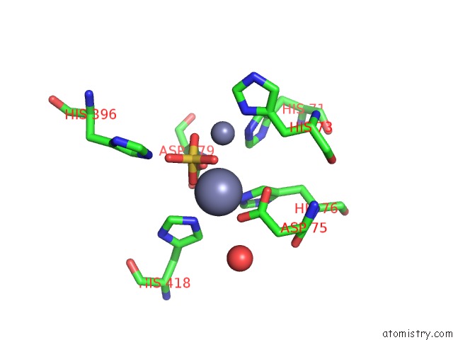
Mono view
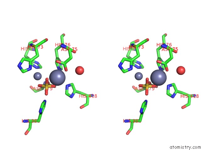
Stereo pair view
Mono view
Stereo pair view
A full contact list of Zinc with other atoms in the Zn binding
site number 1 of Structure of Human Cpsf-73 within 5.0Å range:
|
Zinc binding site 2 out of 2 in 2i7t
Go back to
Zinc binding site 2 out
of 2 in the Structure of Human Cpsf-73
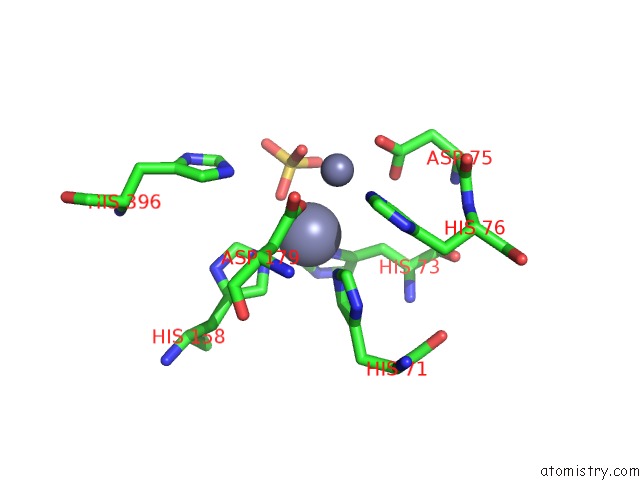
Mono view
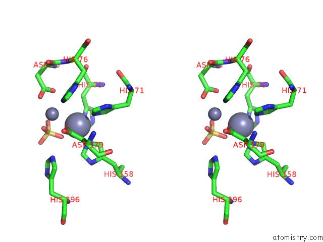
Stereo pair view
Mono view
Stereo pair view
A full contact list of Zinc with other atoms in the Zn binding
site number 2 of Structure of Human Cpsf-73 within 5.0Å range:
|
Reference:
C.R.Mandel,
S.Kaneko,
H.Zhang,
D.Gebauer,
V.Vethantham,
J.L.Manley,
L.Tong.
Polyadenylation Factor Cpsf-73 Is the Pre-Mrna 3'-End-Processing Endonuclease. Nature V. 444 953 2006.
ISSN: ISSN 0028-0836
PubMed: 17128255
DOI: 10.1038/NATURE05363
Page generated: Thu Oct 17 00:51:37 2024
ISSN: ISSN 0028-0836
PubMed: 17128255
DOI: 10.1038/NATURE05363
Last articles
Zn in 9J0NZn in 9J0O
Zn in 9J0P
Zn in 9FJX
Zn in 9EKB
Zn in 9C0F
Zn in 9CAH
Zn in 9CH0
Zn in 9CH3
Zn in 9CH1