Zinc in PDB 2hz8: Qm/Mm Structure Refined From uc(Nmr)-Structure of A Single Chain Diiron Protein
Zinc Binding Sites:
The binding sites of Zinc atom in the Qm/Mm Structure Refined From uc(Nmr)-Structure of A Single Chain Diiron Protein
(pdb code 2hz8). This binding sites where shown within
5.0 Angstroms radius around Zinc atom.
In total 2 binding sites of Zinc where determined in the
Qm/Mm Structure Refined From uc(Nmr)-Structure of A Single Chain Diiron Protein, PDB code: 2hz8:
Jump to Zinc binding site number:
1;
2;
Zinc binding site 1 out
of 2 in 2hz8
Go back to
Zinc Binding Sites List in 2hz8
Zinc binding site 1 out
of 2 in the Qm/Mm Structure Refined From uc(Nmr)-Structure of A Single Chain Diiron Protein
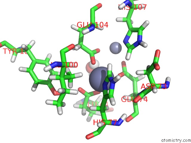
Mono view
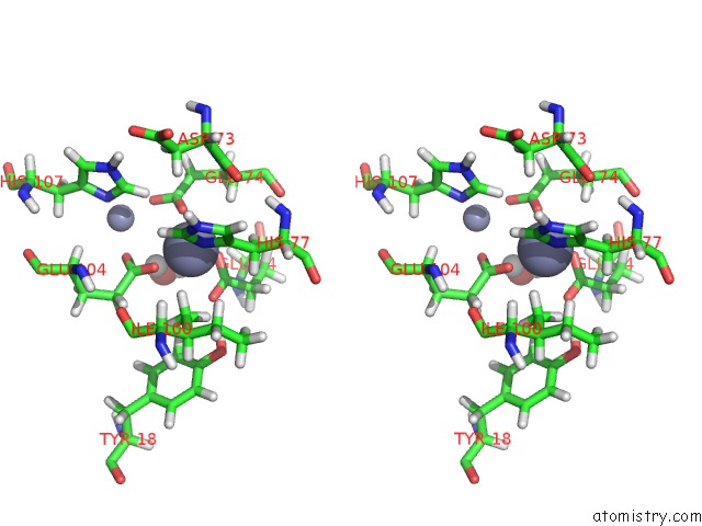
Stereo pair view
|
A full contact list of Zinc with other atoms in the Zn binding
site number 1 of Qm/Mm Structure Refined From uc(Nmr)-Structure of A Single Chain Diiron Protein within 5.0Å range:
probe
|
atom
|
residue
|
distance (Å)
|
B
|
Occ
|
A:Zn116
b:1.0
occ:1.00
|
OE1
|
A:GLU44
|
2.0
|
1.0
|
1.0
|
ND1
|
A:HIS77
|
2.0
|
1.0
|
1.0
|
OE2
|
A:GLU104
|
2.1
|
1.0
|
1.0
|
OE1
|
A:GLU74
|
2.1
|
1.0
|
1.0
|
CD
|
A:GLU44
|
2.7
|
1.0
|
1.0
|
OE2
|
A:GLU44
|
2.7
|
1.0
|
1.0
|
CE1
|
A:HIS77
|
2.9
|
1.0
|
1.0
|
CD
|
A:GLU104
|
3.1
|
1.0
|
1.0
|
HE1
|
A:HIS77
|
3.1
|
1.0
|
1.0
|
CG
|
A:HIS77
|
3.2
|
1.0
|
1.0
|
CD
|
A:GLU74
|
3.2
|
1.0
|
1.0
|
HB2
|
A:HIS77
|
3.2
|
1.0
|
1.0
|
HA
|
A:GLU74
|
3.2
|
0.0
|
1.0
|
OE1
|
A:GLU104
|
3.5
|
1.0
|
1.0
|
HG23
|
A:ILE100
|
3.5
|
0.0
|
1.0
|
CB
|
A:HIS77
|
3.6
|
1.0
|
1.0
|
HB3
|
A:HIS77
|
3.6
|
1.0
|
1.0
|
H1
|
A:HOH118
|
3.6
|
1.0
|
1.0
|
HE1
|
A:HIS107
|
3.7
|
1.0
|
1.0
|
OE2
|
A:GLU74
|
3.8
|
1.0
|
1.0
|
HG22
|
A:ILE100
|
3.9
|
0.0
|
1.0
|
H2
|
A:HOH118
|
3.9
|
1.0
|
1.0
|
NE2
|
A:HIS77
|
4.1
|
1.0
|
1.0
|
CG2
|
A:ILE100
|
4.1
|
0.0
|
1.0
|
CG
|
A:GLU44
|
4.2
|
1.0
|
1.0
|
O
|
A:HOH118
|
4.2
|
1.0
|
1.0
|
ZN
|
A:ZN117
|
4.2
|
1.0
|
1.0
|
HG21
|
A:ILE100
|
4.2
|
0.0
|
1.0
|
CA
|
A:GLU74
|
4.2
|
0.0
|
1.0
|
CD2
|
A:HIS77
|
4.2
|
1.0
|
1.0
|
CG
|
A:GLU74
|
4.3
|
1.0
|
1.0
|
CG
|
A:GLU104
|
4.4
|
1.0
|
1.0
|
HG2
|
A:GLU104
|
4.4
|
1.0
|
1.0
|
HG3
|
A:GLU44
|
4.4
|
1.0
|
1.0
|
HB3
|
A:GLU74
|
4.4
|
1.0
|
1.0
|
HH
|
A:TYR18
|
4.5
|
0.0
|
1.0
|
HA
|
A:GLU44
|
4.5
|
0.0
|
1.0
|
CB
|
A:GLU74
|
4.6
|
1.0
|
1.0
|
CE1
|
A:HIS107
|
4.6
|
1.0
|
1.0
|
HG3
|
A:GLU104
|
4.7
|
1.0
|
1.0
|
O
|
A:ASP73
|
4.7
|
0.0
|
1.0
|
HE2
|
A:TYR18
|
4.7
|
0.0
|
1.0
|
N
|
A:GLU74
|
4.7
|
0.0
|
1.0
|
HB3
|
A:GLU44
|
4.7
|
1.0
|
1.0
|
HB3
|
A:ASP73
|
4.7
|
0.0
|
1.0
|
HG2
|
A:GLU44
|
4.8
|
1.0
|
1.0
|
ND1
|
A:HIS107
|
4.9
|
1.0
|
1.0
|
C
|
A:ASP73
|
4.9
|
0.0
|
1.0
|
CB
|
A:GLU44
|
4.9
|
1.0
|
1.0
|
HG3
|
A:GLU74
|
5.0
|
1.0
|
1.0
|
|
Zinc binding site 2 out
of 2 in 2hz8
Go back to
Zinc Binding Sites List in 2hz8
Zinc binding site 2 out
of 2 in the Qm/Mm Structure Refined From uc(Nmr)-Structure of A Single Chain Diiron Protein
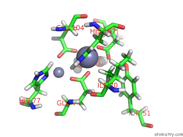
Mono view
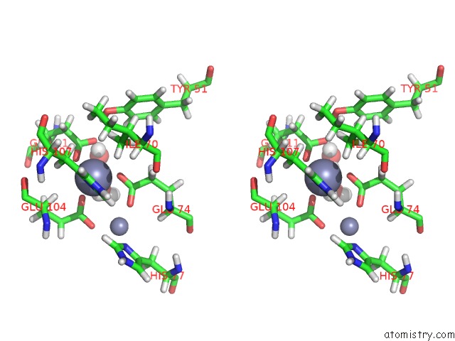
Stereo pair view
|
A full contact list of Zinc with other atoms in the Zn binding
site number 2 of Qm/Mm Structure Refined From uc(Nmr)-Structure of A Single Chain Diiron Protein within 5.0Å range:
probe
|
atom
|
residue
|
distance (Å)
|
B
|
Occ
|
A:Zn117
b:1.0
occ:1.00
|
OE1
|
A:GLU11
|
2.0
|
1.0
|
1.0
|
OE2
|
A:GLU74
|
2.0
|
1.0
|
1.0
|
OE1
|
A:GLU104
|
2.1
|
1.0
|
1.0
|
ND1
|
A:HIS107
|
2.1
|
1.0
|
1.0
|
CD
|
A:GLU11
|
2.9
|
1.0
|
1.0
|
CD
|
A:GLU74
|
3.0
|
1.0
|
1.0
|
CE1
|
A:HIS107
|
3.1
|
1.0
|
1.0
|
CD
|
A:GLU104
|
3.1
|
1.0
|
1.0
|
CG
|
A:HIS107
|
3.1
|
1.0
|
1.0
|
OE2
|
A:GLU11
|
3.2
|
1.0
|
1.0
|
HB2
|
A:HIS107
|
3.2
|
1.0
|
1.0
|
HE1
|
A:HIS107
|
3.3
|
1.0
|
1.0
|
HA
|
A:GLU104
|
3.3
|
0.0
|
1.0
|
OE1
|
A:GLU74
|
3.3
|
1.0
|
1.0
|
H1
|
A:HOH118
|
3.3
|
1.0
|
1.0
|
HB3
|
A:HIS107
|
3.4
|
1.0
|
1.0
|
HG21
|
A:ILE70
|
3.4
|
0.0
|
1.0
|
CB
|
A:HIS107
|
3.5
|
1.0
|
1.0
|
OE2
|
A:GLU104
|
3.6
|
1.0
|
1.0
|
HG23
|
A:ILE70
|
3.8
|
0.0
|
1.0
|
CG2
|
A:ILE70
|
4.0
|
0.0
|
1.0
|
O
|
A:HOH118
|
4.1
|
1.0
|
1.0
|
H1
|
A:HOH119
|
4.1
|
1.0
|
1.0
|
HB3
|
A:GLU104
|
4.2
|
1.0
|
1.0
|
HG22
|
A:ILE70
|
4.2
|
0.0
|
1.0
|
ZN
|
A:ZN116
|
4.2
|
1.0
|
1.0
|
NE2
|
A:HIS107
|
4.2
|
1.0
|
1.0
|
H2
|
A:HOH119
|
4.3
|
1.0
|
1.0
|
CD2
|
A:HIS107
|
4.3
|
1.0
|
1.0
|
CG
|
A:GLU104
|
4.3
|
1.0
|
1.0
|
CG
|
A:GLU11
|
4.3
|
1.0
|
1.0
|
CA
|
A:GLU104
|
4.3
|
0.0
|
1.0
|
CG
|
A:GLU74
|
4.3
|
1.0
|
1.0
|
HG3
|
A:GLU74
|
4.4
|
1.0
|
1.0
|
H2
|
A:HOH118
|
4.4
|
1.0
|
1.0
|
HG3
|
A:GLU11
|
4.4
|
1.0
|
1.0
|
HG2
|
A:GLU74
|
4.5
|
1.0
|
1.0
|
HH
|
A:TYR51
|
4.5
|
0.0
|
1.0
|
CB
|
A:GLU104
|
4.5
|
1.0
|
1.0
|
HA
|
A:GLU11
|
4.5
|
0.0
|
1.0
|
O
|
A:HOH119
|
4.7
|
1.0
|
1.0
|
HB3
|
A:GLU11
|
4.8
|
1.0
|
1.0
|
HG3
|
A:GLU104
|
4.8
|
1.0
|
1.0
|
HE1
|
A:HIS77
|
4.9
|
1.0
|
1.0
|
N
|
A:GLU104
|
5.0
|
0.0
|
1.0
|
|
Reference:
J.R.Calhoun,
W.Liu,
K.Spiegel,
M.Dal Peraro,
M.L.Klein,
K.G.Valentine,
A.J.Wand,
W.F.Degrado.
Solution uc(Nmr) Structure of A Designed Metalloprotein and Complementary Molecular Dynamics Refinement. Structure V. 16 210 2008.
ISSN: ISSN 0969-2126
PubMed: 18275812
DOI: 10.1016/J.STR.2007.11.011
Page generated: Thu Oct 17 00:44:54 2024
|