Zinc »
PDB 2hqk-2ibi »
2hw7 »
Zinc in PDB 2hw7: Crystal Structure of MNK2-D228G in Complex with Staurosporine
Enzymatic activity of Crystal Structure of MNK2-D228G in Complex with Staurosporine
All present enzymatic activity of Crystal Structure of MNK2-D228G in Complex with Staurosporine:
2.7.11.1;
2.7.11.1;
Protein crystallography data
The structure of Crystal Structure of MNK2-D228G in Complex with Staurosporine, PDB code: 2hw7
was solved by
R.Jauch,
M.C.Wahl,
with X-Ray Crystallography technique. A brief refinement statistics is given in the table below:
| Resolution Low / High (Å) | 30.00 / 2.71 |
| Space group | P 32 2 1 |
| Cell size a, b, c (Å), α, β, γ (°) | 102.366, 102.366, 76.439, 90.00, 90.00, 120.00 |
| R / Rfree (%) | 20.1 / 24.6 |
Zinc Binding Sites:
The binding sites of Zinc atom in the Crystal Structure of MNK2-D228G in Complex with Staurosporine
(pdb code 2hw7). This binding sites where shown within
5.0 Angstroms radius around Zinc atom.
In total only one binding site of Zinc was determined in the Crystal Structure of MNK2-D228G in Complex with Staurosporine, PDB code: 2hw7:
In total only one binding site of Zinc was determined in the Crystal Structure of MNK2-D228G in Complex with Staurosporine, PDB code: 2hw7:
Zinc binding site 1 out of 1 in 2hw7
Go back to
Zinc binding site 1 out
of 1 in the Crystal Structure of MNK2-D228G in Complex with Staurosporine
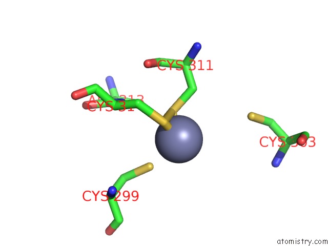
Mono view
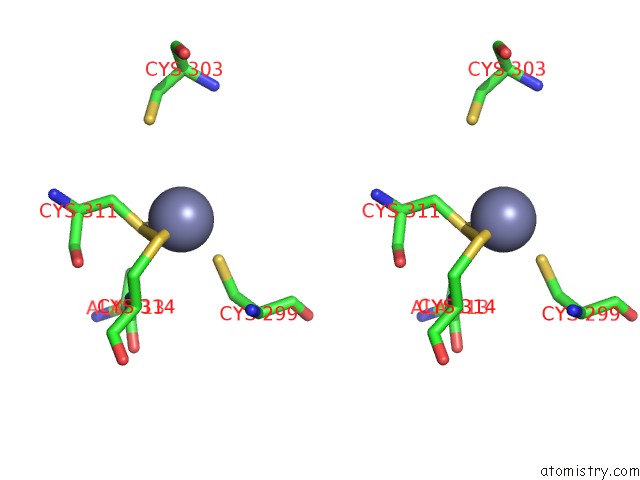
Stereo pair view
Mono view
Stereo pair view
A full contact list of Zinc with other atoms in the Zn binding
site number 1 of Crystal Structure of MNK2-D228G in Complex with Staurosporine within 5.0Å range:
|
Reference:
R.Jauch,
M.K.Cho,
C.Netter,
K.Schreiter,
B.Aicher,
M.Zweckstetter,
M.C.Wahl.
Mitogen-Activated Protein Kinases Interacting Kinases Are Autoinhibited By A Reprogrammed Activation Segment. Embo J. V. 25 4020 2006.
ISSN: ISSN 0261-4189
PubMed: 16917500
DOI: 10.1038/SJ.EMBOJ.7601285
Page generated: Thu Oct 17 00:44:13 2024
ISSN: ISSN 0261-4189
PubMed: 16917500
DOI: 10.1038/SJ.EMBOJ.7601285
Last articles
Zn in 9J0NZn in 9J0O
Zn in 9J0P
Zn in 9FJX
Zn in 9EKB
Zn in 9C0F
Zn in 9CAH
Zn in 9CH0
Zn in 9CH3
Zn in 9CH1