Zinc »
PDB 2fac-2foq »
2ffz »
Zinc in PDB 2ffz: Structural Studies Examining the Substrate Specificity Profiles of Pc- Plcbc Protein Variants
Enzymatic activity of Structural Studies Examining the Substrate Specificity Profiles of Pc- Plcbc Protein Variants
All present enzymatic activity of Structural Studies Examining the Substrate Specificity Profiles of Pc- Plcbc Protein Variants:
3.1.4.3;
3.1.4.3;
Protein crystallography data
The structure of Structural Studies Examining the Substrate Specificity Profiles of Pc- Plcbc Protein Variants, PDB code: 2ffz
was solved by
A.B.Benfield,
N.M.Antikainen,
S.F.Martin,
with X-Ray Crystallography technique. A brief refinement statistics is given in the table below:
| Resolution Low / High (Å) | 25.00 / 2.05 |
| Space group | P 43 21 2 |
| Cell size a, b, c (Å), α, β, γ (°) | 89.396, 89.396, 72.010, 90.00, 90.00, 90.00 |
| R / Rfree (%) | 18 / 21.2 |
Zinc Binding Sites:
The binding sites of Zinc atom in the Structural Studies Examining the Substrate Specificity Profiles of Pc- Plcbc Protein Variants
(pdb code 2ffz). This binding sites where shown within
5.0 Angstroms radius around Zinc atom.
In total 3 binding sites of Zinc where determined in the Structural Studies Examining the Substrate Specificity Profiles of Pc- Plcbc Protein Variants, PDB code: 2ffz:
Jump to Zinc binding site number: 1; 2; 3;
In total 3 binding sites of Zinc where determined in the Structural Studies Examining the Substrate Specificity Profiles of Pc- Plcbc Protein Variants, PDB code: 2ffz:
Jump to Zinc binding site number: 1; 2; 3;
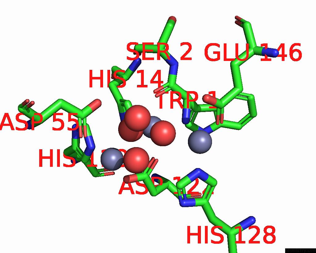
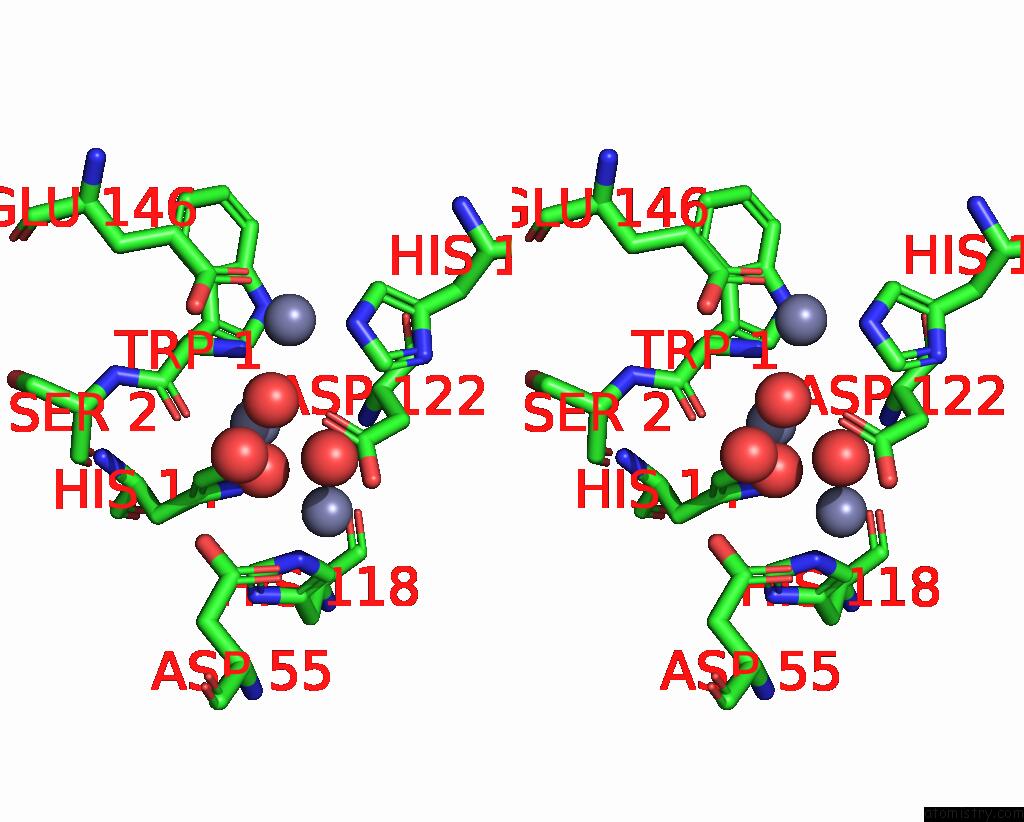
Zinc binding site 1 out of 3 in 2ffz
Go back to
Zinc binding site 1 out
of 3 in the Structural Studies Examining the Substrate Specificity Profiles of Pc- Plcbc Protein Variants
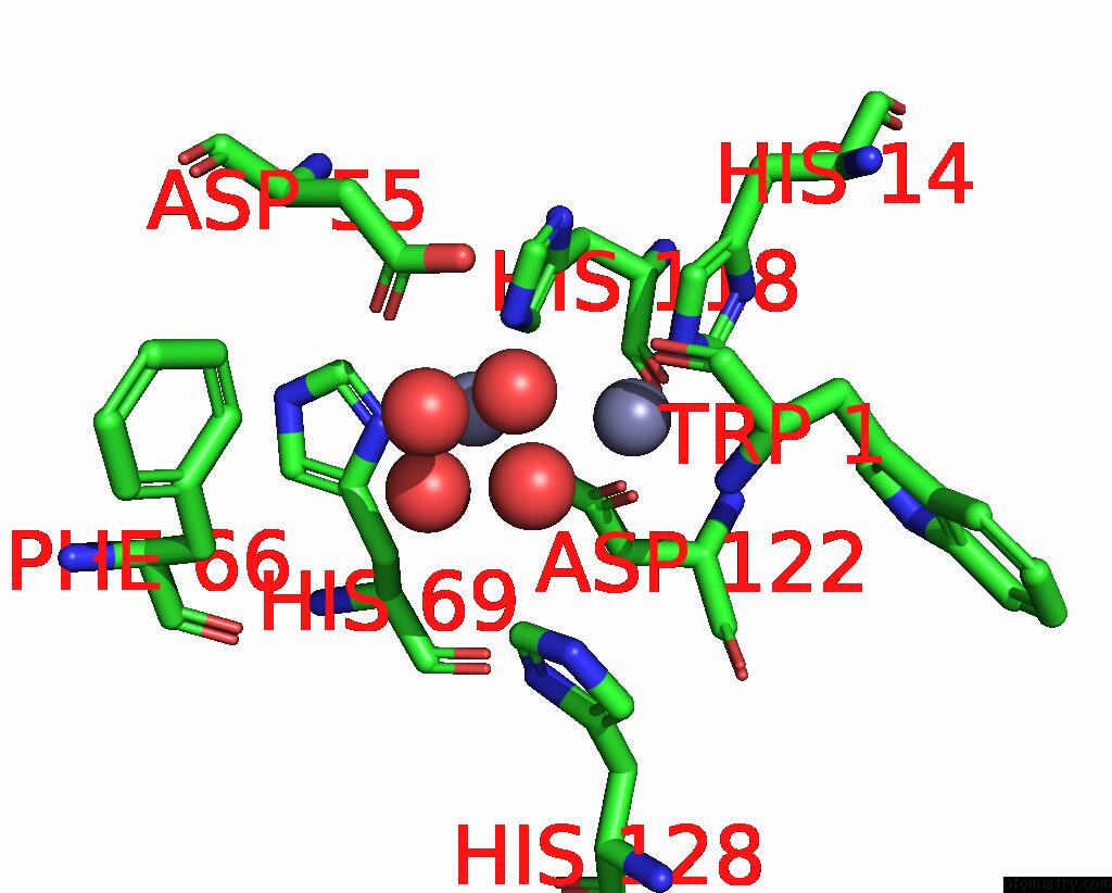
Mono view
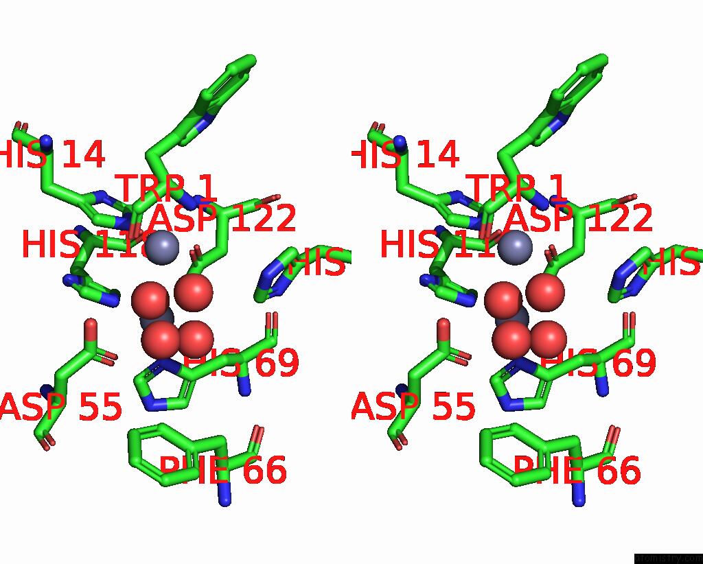
Stereo pair view
Mono view
Stereo pair view
A full contact list of Zinc with other atoms in the Zn binding
site number 1 of Structural Studies Examining the Substrate Specificity Profiles of Pc- Plcbc Protein Variants within 5.0Å range:
|
Zinc binding site 2 out of 3 in 2ffz
Go back to
Zinc binding site 2 out
of 3 in the Structural Studies Examining the Substrate Specificity Profiles of Pc- Plcbc Protein Variants
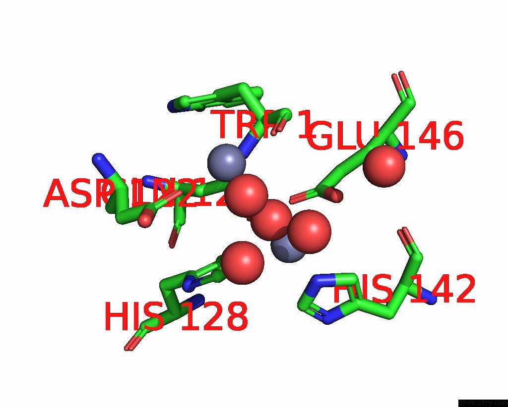
Mono view
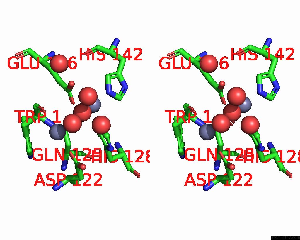
Stereo pair view
Mono view
Stereo pair view
A full contact list of Zinc with other atoms in the Zn binding
site number 2 of Structural Studies Examining the Substrate Specificity Profiles of Pc- Plcbc Protein Variants within 5.0Å range:
|
Zinc binding site 3 out of 3 in 2ffz
Go back to
Zinc binding site 3 out
of 3 in the Structural Studies Examining the Substrate Specificity Profiles of Pc- Plcbc Protein Variants
Mono view
Stereo pair view
Mono view
Stereo pair view
A full contact list of Zinc with other atoms in the Zn binding
site number 3 of Structural Studies Examining the Substrate Specificity Profiles of Pc- Plcbc Protein Variants within 5.0Å range:
|
Reference:
A.P.Benfield,
N.M.Goodey,
L.T.Phillips,
S.F.Martin.
Structural Studies Examining the Substrate Specificity Profiles of Pc-Plc(Bc) Protein Variants. Arch.Biochem.Biophys. V. 460 41 2007.
ISSN: ISSN 0003-9861
PubMed: 17324372
DOI: 10.1016/J.ABB.2007.01.023
Page generated: Wed Oct 16 23:45:46 2024
ISSN: ISSN 0003-9861
PubMed: 17324372
DOI: 10.1016/J.ABB.2007.01.023
Last articles
Zn in 9J0NZn in 9J0O
Zn in 9J0P
Zn in 9FJX
Zn in 9EKB
Zn in 9C0F
Zn in 9CAH
Zn in 9CH0
Zn in 9CH3
Zn in 9CH1