Zinc »
PDB 2fac-2foq »
2fad »
Zinc in PDB 2fad: Crystal Structure of E. Coli Heptanoyl-Acp
Protein crystallography data
The structure of Crystal Structure of E. Coli Heptanoyl-Acp, PDB code: 2fad
was solved by
A.Roujeinikova,
with X-Ray Crystallography technique. A brief refinement statistics is given in the table below:
| Resolution Low / High (Å) | 10.00 / 1.60 |
| Space group | P 21 21 2 |
| Cell size a, b, c (Å), α, β, γ (°) | 49.257, 106.244, 28.195, 90.00, 90.00, 90.00 |
| R / Rfree (%) | 20.9 / 25.6 |
Other elements in 2fad:
The structure of Crystal Structure of E. Coli Heptanoyl-Acp also contains other interesting chemical elements:
| Sodium | (Na) | 1 atom |
Zinc Binding Sites:
The binding sites of Zinc atom in the Crystal Structure of E. Coli Heptanoyl-Acp
(pdb code 2fad). This binding sites where shown within
5.0 Angstroms radius around Zinc atom.
In total 8 binding sites of Zinc where determined in the Crystal Structure of E. Coli Heptanoyl-Acp, PDB code: 2fad:
Jump to Zinc binding site number: 1; 2; 3; 4; 5; 6; 7; 8;
In total 8 binding sites of Zinc where determined in the Crystal Structure of E. Coli Heptanoyl-Acp, PDB code: 2fad:
Jump to Zinc binding site number: 1; 2; 3; 4; 5; 6; 7; 8;
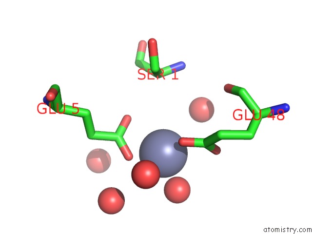
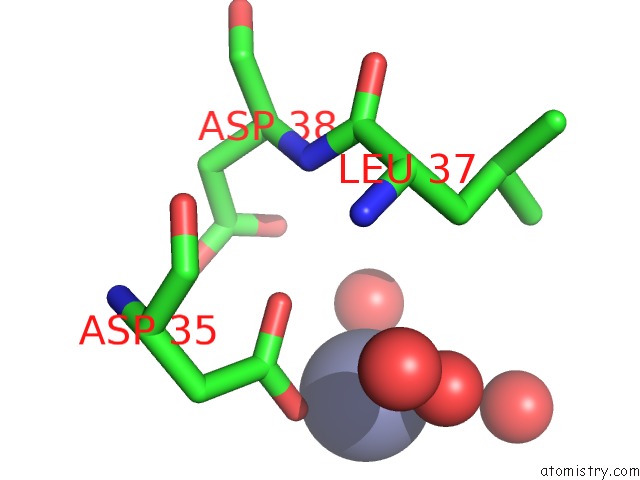
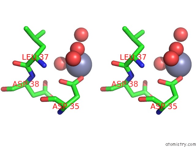
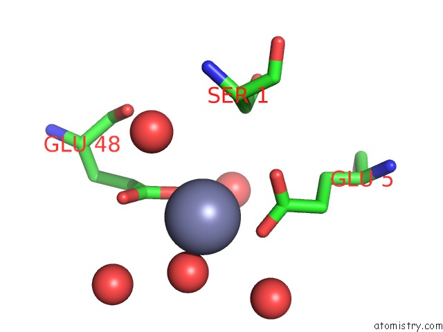
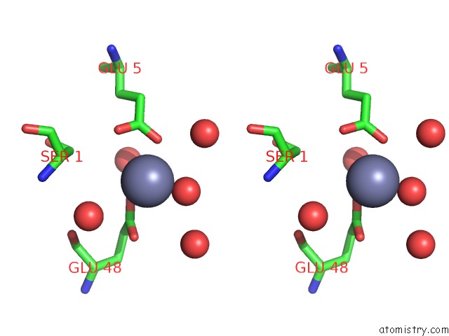
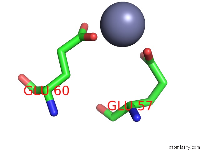
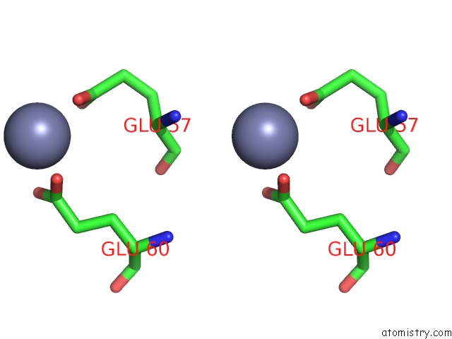
Zinc binding site 1 out of 8 in 2fad
Go back to
Zinc binding site 1 out
of 8 in the Crystal Structure of E. Coli Heptanoyl-Acp
Mono view
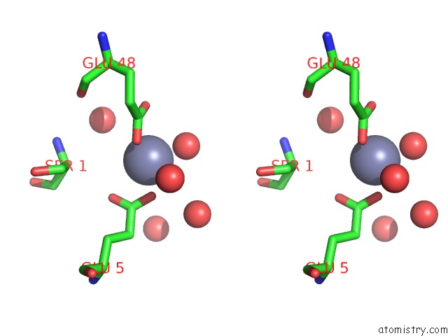
Stereo pair view
Mono view
Stereo pair view
A full contact list of Zinc with other atoms in the Zn binding
site number 1 of Crystal Structure of E. Coli Heptanoyl-Acp within 5.0Å range:
|
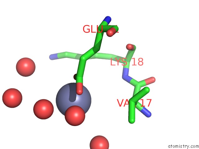
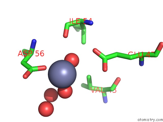
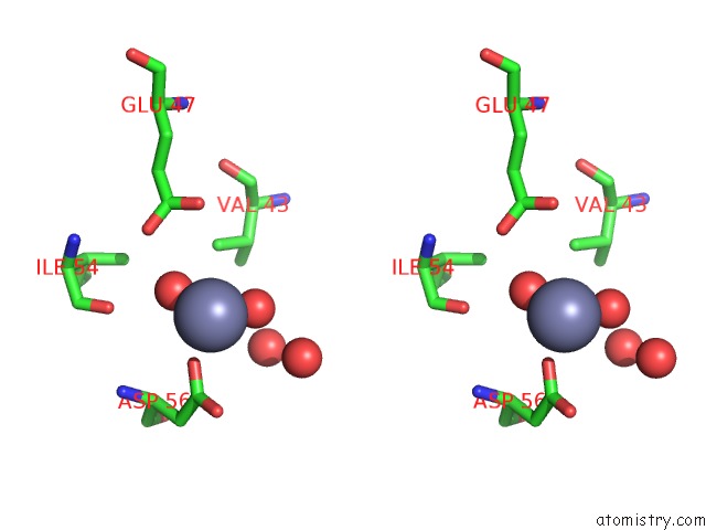
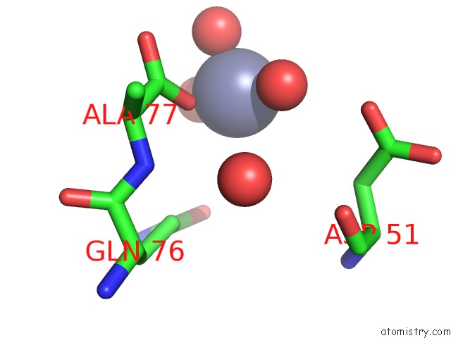
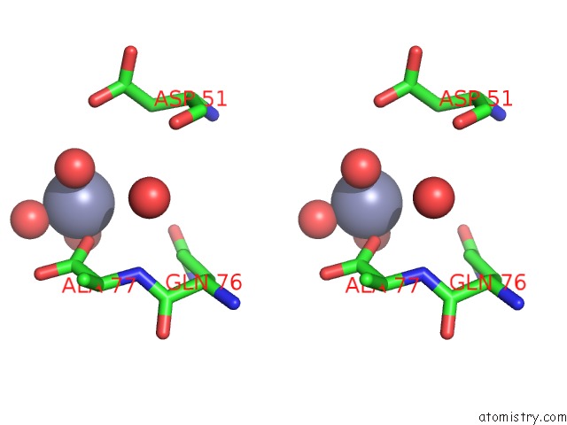
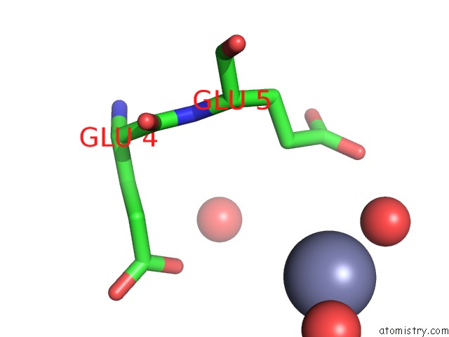
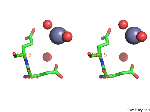
Zinc binding site 2 out of 8 in 2fad
Go back to
Zinc binding site 2 out
of 8 in the Crystal Structure of E. Coli Heptanoyl-Acp
Mono view
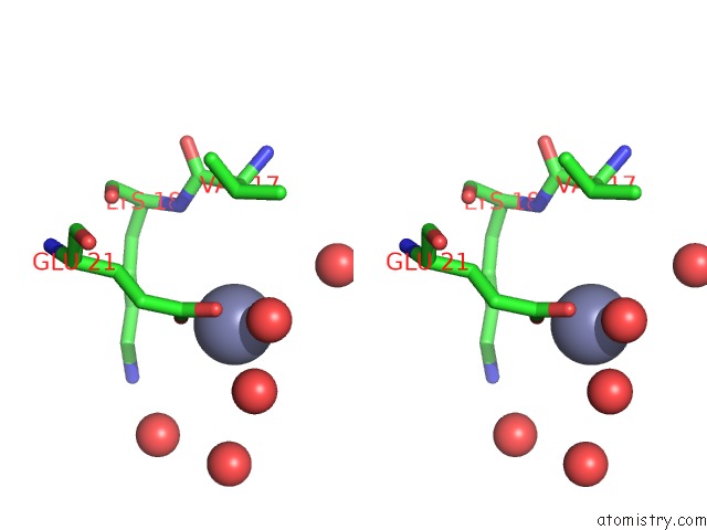
Stereo pair view
Mono view
Stereo pair view
A full contact list of Zinc with other atoms in the Zn binding
site number 2 of Crystal Structure of E. Coli Heptanoyl-Acp within 5.0Å range:
|
Zinc binding site 3 out of 8 in 2fad
Go back to
Zinc binding site 3 out
of 8 in the Crystal Structure of E. Coli Heptanoyl-Acp
Mono view
Stereo pair view
Mono view
Stereo pair view
A full contact list of Zinc with other atoms in the Zn binding
site number 3 of Crystal Structure of E. Coli Heptanoyl-Acp within 5.0Å range:
|
Zinc binding site 4 out of 8 in 2fad
Go back to
Zinc binding site 4 out
of 8 in the Crystal Structure of E. Coli Heptanoyl-Acp
Mono view
Stereo pair view
Mono view
Stereo pair view
A full contact list of Zinc with other atoms in the Zn binding
site number 4 of Crystal Structure of E. Coli Heptanoyl-Acp within 5.0Å range:
|
Zinc binding site 5 out of 8 in 2fad
Go back to
Zinc binding site 5 out
of 8 in the Crystal Structure of E. Coli Heptanoyl-Acp
Mono view
Stereo pair view
Mono view
Stereo pair view
A full contact list of Zinc with other atoms in the Zn binding
site number 5 of Crystal Structure of E. Coli Heptanoyl-Acp within 5.0Å range:
|
Zinc binding site 6 out of 8 in 2fad
Go back to
Zinc binding site 6 out
of 8 in the Crystal Structure of E. Coli Heptanoyl-Acp
Mono view
Stereo pair view
Mono view
Stereo pair view
A full contact list of Zinc with other atoms in the Zn binding
site number 6 of Crystal Structure of E. Coli Heptanoyl-Acp within 5.0Å range:
|
Zinc binding site 7 out of 8 in 2fad
Go back to
Zinc binding site 7 out
of 8 in the Crystal Structure of E. Coli Heptanoyl-Acp
Mono view
Stereo pair view
Mono view
Stereo pair view
A full contact list of Zinc with other atoms in the Zn binding
site number 7 of Crystal Structure of E. Coli Heptanoyl-Acp within 5.0Å range:
|
Zinc binding site 8 out of 8 in 2fad
Go back to
Zinc binding site 8 out
of 8 in the Crystal Structure of E. Coli Heptanoyl-Acp
Mono view
Stereo pair view
Mono view
Stereo pair view
A full contact list of Zinc with other atoms in the Zn binding
site number 8 of Crystal Structure of E. Coli Heptanoyl-Acp within 5.0Å range:
|
Reference:
A.Roujeinikova,
W.J.Simon,
J.Gilroy,
D.W.Rice,
J.B.Rafferty,
A.R.Slabas.
Structural Studies of Fatty Acyl-(Acyl Carrier Protein) Thioesters Reveal A Hydrophobic Binding Cavity That Can Expand to Fit Longer Substrates. J.Mol.Biol. V. 365 135 2007.
ISSN: ISSN 0022-2836
PubMed: 17059829
DOI: 10.1016/J.JMB.2006.09.049
Page generated: Wed Oct 16 23:42:15 2024
ISSN: ISSN 0022-2836
PubMed: 17059829
DOI: 10.1016/J.JMB.2006.09.049
Last articles
Zn in 9J0NZn in 9J0O
Zn in 9J0P
Zn in 9FJX
Zn in 9EKB
Zn in 9C0F
Zn in 9CAH
Zn in 9CH0
Zn in 9CH3
Zn in 9CH1