Zinc »
PDB 2e3x-2eer »
2e47 »
Zinc in PDB 2e47: Crystal Structure Analysis of the Clock Protein EA4 (Glycosylation Form)
Protein crystallography data
The structure of Crystal Structure Analysis of the Clock Protein EA4 (Glycosylation Form), PDB code: 2e47
was solved by
S.-Y.Park,
T.Hiraki,
with X-Ray Crystallography technique. A brief refinement statistics is given in the table below:
| Resolution Low / High (Å) | 20.00 / 2.11 |
| Space group | P 1 21 1 |
| Cell size a, b, c (Å), α, β, γ (°) | 47.098, 73.894, 47.446, 90.00, 104.07, 90.00 |
| R / Rfree (%) | 17 / 25.1 |
Other elements in 2e47:
The structure of Crystal Structure Analysis of the Clock Protein EA4 (Glycosylation Form) also contains other interesting chemical elements:
| Copper | (Cu) | 2 atoms |
Zinc Binding Sites:
The binding sites of Zinc atom in the Crystal Structure Analysis of the Clock Protein EA4 (Glycosylation Form)
(pdb code 2e47). This binding sites where shown within
5.0 Angstroms radius around Zinc atom.
In total 2 binding sites of Zinc where determined in the Crystal Structure Analysis of the Clock Protein EA4 (Glycosylation Form), PDB code: 2e47:
Jump to Zinc binding site number: 1; 2;
In total 2 binding sites of Zinc where determined in the Crystal Structure Analysis of the Clock Protein EA4 (Glycosylation Form), PDB code: 2e47:
Jump to Zinc binding site number: 1; 2;
Zinc binding site 1 out of 2 in 2e47
Go back to
Zinc binding site 1 out
of 2 in the Crystal Structure Analysis of the Clock Protein EA4 (Glycosylation Form)
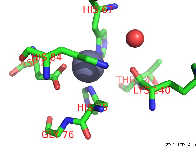
Mono view
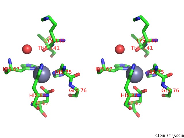
Stereo pair view
Mono view
Stereo pair view
A full contact list of Zinc with other atoms in the Zn binding
site number 1 of Crystal Structure Analysis of the Clock Protein EA4 (Glycosylation Form) within 5.0Å range:
|
Zinc binding site 2 out of 2 in 2e47
Go back to
Zinc binding site 2 out
of 2 in the Crystal Structure Analysis of the Clock Protein EA4 (Glycosylation Form)
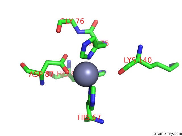
Mono view
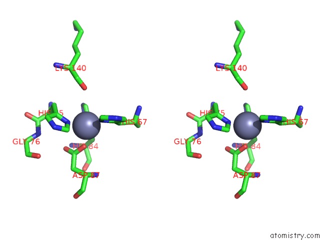
Stereo pair view
Mono view
Stereo pair view
A full contact list of Zinc with other atoms in the Zn binding
site number 2 of Crystal Structure Analysis of the Clock Protein EA4 (Glycosylation Form) within 5.0Å range:
|
Reference:
T.Hiraki,
N.Shibayama,
J.R.M.Tame,
S.Akashi,
S.-Y.Park.
The Clock Protein EA4 Ticks Away with Movement of A Mobile Copper Ion To Be Published.
Page generated: Wed Oct 16 22:58:44 2024
Last articles
Zn in 9J0NZn in 9J0O
Zn in 9J0P
Zn in 9FJX
Zn in 9EKB
Zn in 9C0F
Zn in 9CAH
Zn in 9CH0
Zn in 9CH3
Zn in 9CH1