Zinc »
PDB 2ds5-2e2z »
2ds5 »
Zinc in PDB 2ds5: Structure of the Zbd in the Orthorhomibic Crystal From
Protein crystallography data
The structure of Structure of the Zbd in the Orthorhomibic Crystal From, PDB code: 2ds5
was solved by
H.K.Song,
E.Y.Park,
B.G.Lee,
S.B.Hong,
with X-Ray Crystallography technique. A brief refinement statistics is given in the table below:
| Resolution Low / High (Å) | 50.00 / 1.50 |
| Space group | P 21 21 21 |
| Cell size a, b, c (Å), α, β, γ (°) | 32.523, 43.399, 67.719, 90.00, 90.00, 90.00 |
| R / Rfree (%) | 18.5 / 22.1 |
Other elements in 2ds5:
The structure of Structure of the Zbd in the Orthorhomibic Crystal From also contains other interesting chemical elements:
| Calcium | (Ca) | 1 atom |
Zinc Binding Sites:
The binding sites of Zinc atom in the Structure of the Zbd in the Orthorhomibic Crystal From
(pdb code 2ds5). This binding sites where shown within
5.0 Angstroms radius around Zinc atom.
In total 2 binding sites of Zinc where determined in the Structure of the Zbd in the Orthorhomibic Crystal From, PDB code: 2ds5:
Jump to Zinc binding site number: 1; 2;
In total 2 binding sites of Zinc where determined in the Structure of the Zbd in the Orthorhomibic Crystal From, PDB code: 2ds5:
Jump to Zinc binding site number: 1; 2;
Zinc binding site 1 out of 2 in 2ds5
Go back to
Zinc binding site 1 out
of 2 in the Structure of the Zbd in the Orthorhomibic Crystal From
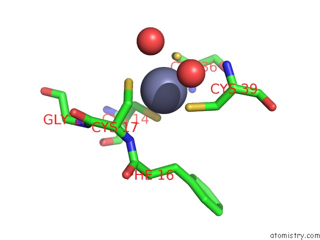
Mono view
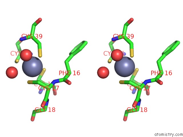
Stereo pair view
Mono view
Stereo pair view
A full contact list of Zinc with other atoms in the Zn binding
site number 1 of Structure of the Zbd in the Orthorhomibic Crystal From within 5.0Å range:
|
Zinc binding site 2 out of 2 in 2ds5
Go back to
Zinc binding site 2 out
of 2 in the Structure of the Zbd in the Orthorhomibic Crystal From
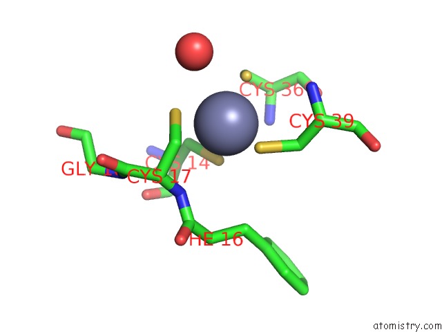
Mono view
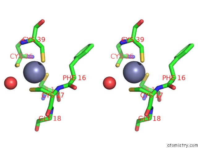
Stereo pair view
Mono view
Stereo pair view
A full contact list of Zinc with other atoms in the Zn binding
site number 2 of Structure of the Zbd in the Orthorhomibic Crystal From within 5.0Å range:
|
Reference:
E.Y.Park,
B.G.Lee,
S.B.Hong,
H.W.Kim,
H.Jeon,
H.K.Song.
Structural Basis of Sspb-Tail Recognition By the Zinc Binding Domain of Clpx. J.Mol.Biol. V. 367 514 2007.
ISSN: ISSN 0022-2836
PubMed: 17258768
DOI: 10.1016/J.JMB.2007.01.003
Page generated: Wed Oct 16 22:50:50 2024
ISSN: ISSN 0022-2836
PubMed: 17258768
DOI: 10.1016/J.JMB.2007.01.003
Last articles
Zn in 9J0NZn in 9J0O
Zn in 9J0P
Zn in 9FJX
Zn in 9EKB
Zn in 9C0F
Zn in 9CAH
Zn in 9CH0
Zn in 9CH3
Zn in 9CH1