Zinc »
PDB 2db6-2drp »
2dq6 »
Zinc in PDB 2dq6: Crystal Structure of Aminopeptidase N From Escherichia Coli
Enzymatic activity of Crystal Structure of Aminopeptidase N From Escherichia Coli
All present enzymatic activity of Crystal Structure of Aminopeptidase N From Escherichia Coli:
3.4.11.2;
3.4.11.2;
Protein crystallography data
The structure of Crystal Structure of Aminopeptidase N From Escherichia Coli, PDB code: 2dq6
was solved by
Y.Nakajima,
Y.Onohara,
K.Ito,
T.Yoshimoto,
with X-Ray Crystallography technique. A brief refinement statistics is given in the table below:
| Resolution Low / High (Å) | 20.00 / 1.50 |
| Space group | P 31 2 1 |
| Cell size a, b, c (Å), α, β, γ (°) | 120.480, 120.480, 170.791, 90.00, 90.00, 120.00 |
| R / Rfree (%) | 18.1 / 19.1 |
Zinc Binding Sites:
The binding sites of Zinc atom in the Crystal Structure of Aminopeptidase N From Escherichia Coli
(pdb code 2dq6). This binding sites where shown within
5.0 Angstroms radius around Zinc atom.
In total only one binding site of Zinc was determined in the Crystal Structure of Aminopeptidase N From Escherichia Coli, PDB code: 2dq6:
In total only one binding site of Zinc was determined in the Crystal Structure of Aminopeptidase N From Escherichia Coli, PDB code: 2dq6:
Zinc binding site 1 out of 1 in 2dq6
Go back to
Zinc binding site 1 out
of 1 in the Crystal Structure of Aminopeptidase N From Escherichia Coli
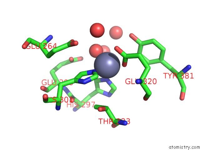
Mono view
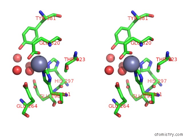
Stereo pair view
Mono view
Stereo pair view
A full contact list of Zinc with other atoms in the Zn binding
site number 1 of Crystal Structure of Aminopeptidase N From Escherichia Coli within 5.0Å range:
|
Reference:
K.Ito,
Y.Nakajima,
Y.Onohara,
M.Takeo,
K.Nakashima,
F.Matsubara,
T.Ito,
T.Yoshimoto.
Aminopeptidase N (Proteobacteria Alanyl Aminopeptidase) From Escherichia Coli: Crystal Structure and Conformational Change of the Methionine 260 Residue Involved in Substrate Recognition J.Biol.Chem. V. 281 33664 2006.
ISSN: ISSN 0021-9258
PubMed: 16885166
DOI: 10.1074/JBC.M605203200
Page generated: Wed Oct 16 22:50:33 2024
ISSN: ISSN 0021-9258
PubMed: 16885166
DOI: 10.1074/JBC.M605203200
Last articles
Zn in 9J0NZn in 9J0O
Zn in 9J0P
Zn in 9FJX
Zn in 9EKB
Zn in 9C0F
Zn in 9CAH
Zn in 9CH0
Zn in 9CH3
Zn in 9CH1