Zinc »
PDB 2c7a-2cij »
2cey »
Zinc in PDB 2cey: Apo Structure of Siap
Protein crystallography data
The structure of Apo Structure of Siap, PDB code: 2cey
was solved by
A.Muller,
E.Severi,
C.Mulligan,
A.G.Watts,
D.J.Kelly,
K.S.Wilson,
A.J.Wilkinson,
G.H.Thomas,
with X-Ray Crystallography technique. A brief refinement statistics is given in the table below:
| Resolution Low / High (Å) | 101.53 / 1.70 |
| Space group | I 2 2 2 |
| Cell size a, b, c (Å), α, β, γ (°) | 46.761, 102.508, 202.648, 90.00, 90.00, 90.00 |
| R / Rfree (%) | 19.4 / 23.6 |
Zinc Binding Sites:
The binding sites of Zinc atom in the Apo Structure of Siap
(pdb code 2cey). This binding sites where shown within
5.0 Angstroms radius around Zinc atom.
In total 3 binding sites of Zinc where determined in the Apo Structure of Siap, PDB code: 2cey:
Jump to Zinc binding site number: 1; 2; 3;
In total 3 binding sites of Zinc where determined in the Apo Structure of Siap, PDB code: 2cey:
Jump to Zinc binding site number: 1; 2; 3;
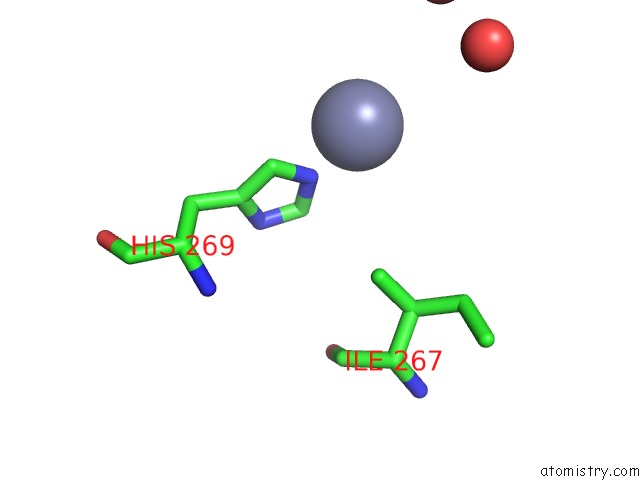
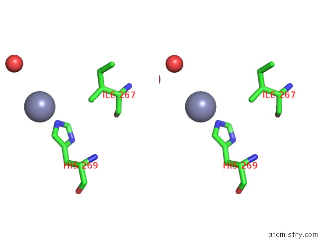
Zinc binding site 1 out of 3 in 2cey
Go back to
Zinc binding site 1 out
of 3 in the Apo Structure of Siap
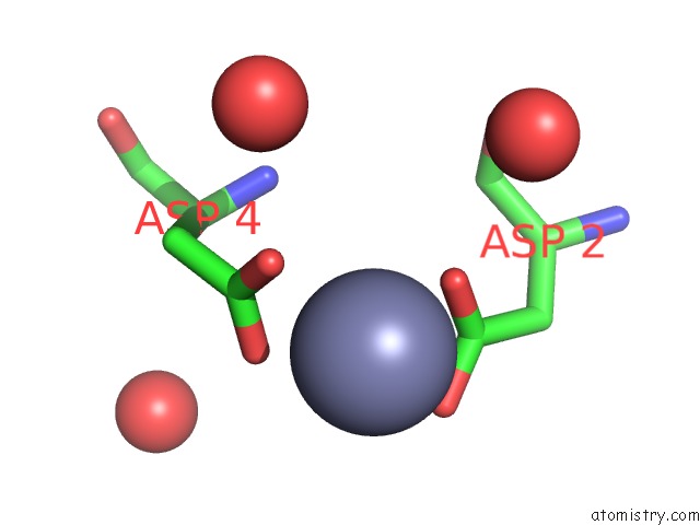
Mono view
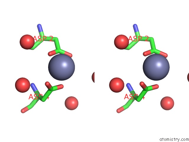
Stereo pair view
Mono view
Stereo pair view
A full contact list of Zinc with other atoms in the Zn binding
site number 1 of Apo Structure of Siap within 5.0Å range:
|
Zinc binding site 2 out of 3 in 2cey
Go back to
Zinc binding site 2 out
of 3 in the Apo Structure of Siap
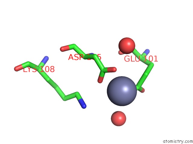
Mono view
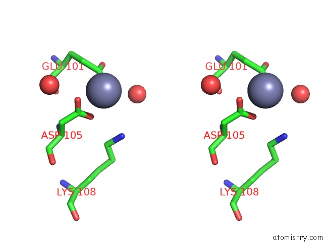
Stereo pair view
Mono view
Stereo pair view
A full contact list of Zinc with other atoms in the Zn binding
site number 2 of Apo Structure of Siap within 5.0Å range:
|
Zinc binding site 3 out of 3 in 2cey
Go back to
Zinc binding site 3 out
of 3 in the Apo Structure of Siap
Mono view
Stereo pair view
Mono view
Stereo pair view
A full contact list of Zinc with other atoms in the Zn binding
site number 3 of Apo Structure of Siap within 5.0Å range:
|
Reference:
A.Muller,
E.Severi,
C.Mulligan,
A.G.Watts,
D.J.Kelly,
K.S.Wilson,
A.J.Wilkinson,
G.H.Thomas.
Conservation of Structure and Mechanism in Primary and Secondary Transporters Exemplified By Siap, A Sialic Acid Binding Virulence Factor From Haemophilus Influenzae J.Biol.Chem. V. 281 22212 2006.
ISSN: ISSN 0021-9258
PubMed: 16702222
DOI: 10.1074/JBC.M603463200
Page generated: Wed Oct 16 22:20:31 2024
ISSN: ISSN 0021-9258
PubMed: 16702222
DOI: 10.1074/JBC.M603463200
Last articles
Zn in 9J0NZn in 9J0O
Zn in 9J0P
Zn in 9FJX
Zn in 9EKB
Zn in 9C0F
Zn in 9CAH
Zn in 9CH0
Zn in 9CH3
Zn in 9CH1