Zinc »
PDB 2c7a-2cij »
2cbn »
Zinc in PDB 2cbn: Crystal Structure of Zipd From Escherichia Coli
Enzymatic activity of Crystal Structure of Zipd From Escherichia Coli
All present enzymatic activity of Crystal Structure of Zipd From Escherichia Coli:
3.1.26.11;
3.1.26.11;
Protein crystallography data
The structure of Crystal Structure of Zipd From Escherichia Coli, PDB code: 2cbn
was solved by
E.Pohl,
W.Meyer-Klaucke,
B.Kostelecky,
with X-Ray Crystallography technique. A brief refinement statistics is given in the table below:
| Resolution Low / High (Å) | 50 / 2.9 |
| Space group | P 64 2 2 |
| Cell size a, b, c (Å), α, β, γ (°) | 147.200, 147.200, 138.200, 90.00, 90.00, 120.00 |
| R / Rfree (%) | 23 / 25 |
Zinc Binding Sites:
The binding sites of Zinc atom in the Crystal Structure of Zipd From Escherichia Coli
(pdb code 2cbn). This binding sites where shown within
5.0 Angstroms radius around Zinc atom.
In total 2 binding sites of Zinc where determined in the Crystal Structure of Zipd From Escherichia Coli, PDB code: 2cbn:
Jump to Zinc binding site number: 1; 2;
In total 2 binding sites of Zinc where determined in the Crystal Structure of Zipd From Escherichia Coli, PDB code: 2cbn:
Jump to Zinc binding site number: 1; 2;
Zinc binding site 1 out of 2 in 2cbn
Go back to
Zinc binding site 1 out
of 2 in the Crystal Structure of Zipd From Escherichia Coli
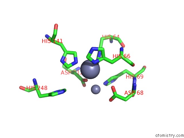
Mono view
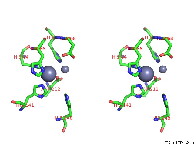
Stereo pair view
Mono view
Stereo pair view
A full contact list of Zinc with other atoms in the Zn binding
site number 1 of Crystal Structure of Zipd From Escherichia Coli within 5.0Å range:
|
Zinc binding site 2 out of 2 in 2cbn
Go back to
Zinc binding site 2 out
of 2 in the Crystal Structure of Zipd From Escherichia Coli
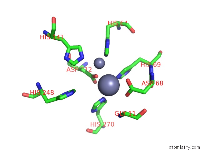
Mono view
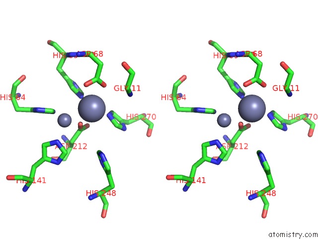
Stereo pair view
Mono view
Stereo pair view
A full contact list of Zinc with other atoms in the Zn binding
site number 2 of Crystal Structure of Zipd From Escherichia Coli within 5.0Å range:
|
Reference:
B.Kostelecky,
E.Pohl,
A.Vogel,
O.Schilling,
W.Meyer-Klaucke.
The Crystal Structure of the Zinc Phosphodiesterase From Escherichia Coli Provides Insight Into Function and Cooperativity of Trnase Z-Family Proteins. J.Bacteriol. V. 188 1607 2006.
ISSN: ISSN 0021-9193
PubMed: 16452444
DOI: 10.1128/JB.188.4.1607-1614.2006
Page generated: Wed Oct 16 22:16:34 2024
ISSN: ISSN 0021-9193
PubMed: 16452444
DOI: 10.1128/JB.188.4.1607-1614.2006
Last articles
Zn in 9J0NZn in 9J0O
Zn in 9J0P
Zn in 9FJX
Zn in 9EKB
Zn in 9C0F
Zn in 9CAH
Zn in 9CH0
Zn in 9CH3
Zn in 9CH1