Zinc »
PDB 2b8k-2bnm »
2bin »
Zinc in PDB 2bin: Human P53 Core Domain Mutant M133L-H168R-V203A-N239Y-N268D
Protein crystallography data
The structure of Human P53 Core Domain Mutant M133L-H168R-V203A-N239Y-N268D, PDB code: 2bin
was solved by
A.C.Joerger,
A.R.Fersht,
with X-Ray Crystallography technique. A brief refinement statistics is given in the table below:
| Resolution Low / High (Å) | 55.0 / 1.9 |
| Space group | P 65 2 2 |
| Cell size a, b, c (Å), α, β, γ (°) | 45.045, 45.045, 331.780, 90.00, 90.00, 120.00 |
| R / Rfree (%) | 19.9 / 23.6 |
Zinc Binding Sites:
The binding sites of Zinc atom in the Human P53 Core Domain Mutant M133L-H168R-V203A-N239Y-N268D
(pdb code 2bin). This binding sites where shown within
5.0 Angstroms radius around Zinc atom.
In total only one binding site of Zinc was determined in the Human P53 Core Domain Mutant M133L-H168R-V203A-N239Y-N268D, PDB code: 2bin:
In total only one binding site of Zinc was determined in the Human P53 Core Domain Mutant M133L-H168R-V203A-N239Y-N268D, PDB code: 2bin:
Zinc binding site 1 out of 1 in 2bin
Go back to
Zinc binding site 1 out
of 1 in the Human P53 Core Domain Mutant M133L-H168R-V203A-N239Y-N268D
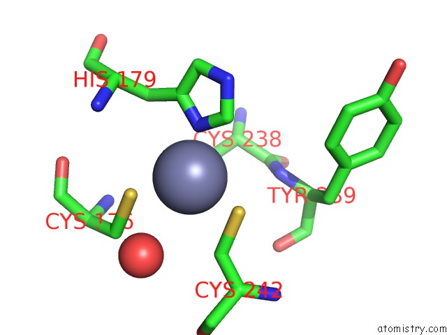
Mono view
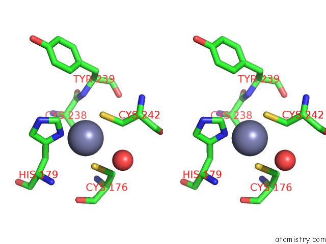
Stereo pair view
Mono view
Stereo pair view
A full contact list of Zinc with other atoms in the Zn binding
site number 1 of Human P53 Core Domain Mutant M133L-H168R-V203A-N239Y-N268D within 5.0Å range:
|
Reference:
A.C.Joerger,
H.C.Ang,
D.B.Veprintsev,
C.M.Blair,
A.R.Fersht.
Structures of P53 Cancer Mutants and Mechanism of Rescue By Second-Site Suppressor Mutations J.Biol.Chem. V. 280 16030 2005.
ISSN: ISSN 0021-9258
PubMed: 15703170
DOI: 10.1074/JBC.M500179200
Page generated: Wed Oct 16 22:02:56 2024
ISSN: ISSN 0021-9258
PubMed: 15703170
DOI: 10.1074/JBC.M500179200
Last articles
Zn in 9MJ5Zn in 9HNW
Zn in 9G0L
Zn in 9FNE
Zn in 9DZN
Zn in 9E0I
Zn in 9D32
Zn in 9DAK
Zn in 8ZXC
Zn in 8ZUF