Zinc »
PDB 2afo-2aqr »
2aq2 »
Zinc in PDB 2aq2: Crystal Structure of T-Cell Receptor V Beta Domain Variant Complexed with Superantigen SEC3 Mutant
Protein crystallography data
The structure of Crystal Structure of T-Cell Receptor V Beta Domain Variant Complexed with Superantigen SEC3 Mutant, PDB code: 2aq2
was solved by
S.Cho,
C.P.Swaminathan,
J.Yang,
M.C.Kerzic,
R.Guan,
M.C.Kieke,
D.M.Kranz,
R.A.Mariuzza,
E.J.Sundberg,
with X-Ray Crystallography technique. A brief refinement statistics is given in the table below:
| Resolution Low / High (Å) | 40.00 / 1.80 |
| Space group | P 65 |
| Cell size a, b, c (Å), α, β, γ (°) | 96.537, 96.537, 92.182, 90.00, 90.00, 120.00 |
| R / Rfree (%) | 18.5 / 21.3 |
Other elements in 2aq2:
The structure of Crystal Structure of T-Cell Receptor V Beta Domain Variant Complexed with Superantigen SEC3 Mutant also contains other interesting chemical elements:
| Sodium | (Na) | 3 atoms |
Zinc Binding Sites:
The binding sites of Zinc atom in the Crystal Structure of T-Cell Receptor V Beta Domain Variant Complexed with Superantigen SEC3 Mutant
(pdb code 2aq2). This binding sites where shown within
5.0 Angstroms radius around Zinc atom.
In total only one binding site of Zinc was determined in the Crystal Structure of T-Cell Receptor V Beta Domain Variant Complexed with Superantigen SEC3 Mutant, PDB code: 2aq2:
In total only one binding site of Zinc was determined in the Crystal Structure of T-Cell Receptor V Beta Domain Variant Complexed with Superantigen SEC3 Mutant, PDB code: 2aq2:
Zinc binding site 1 out of 1 in 2aq2
Go back to
Zinc binding site 1 out
of 1 in the Crystal Structure of T-Cell Receptor V Beta Domain Variant Complexed with Superantigen SEC3 Mutant
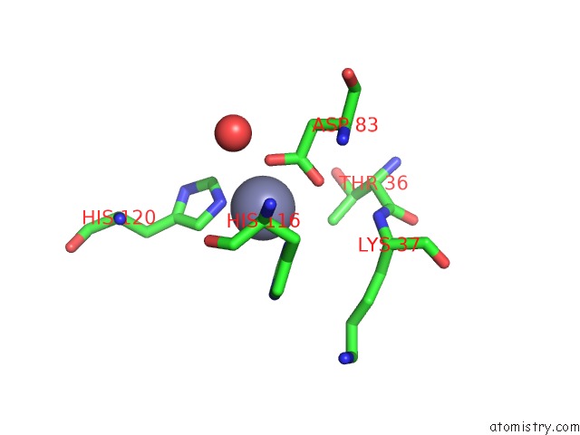
Mono view
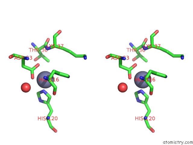
Stereo pair view
Mono view
Stereo pair view
A full contact list of Zinc with other atoms in the Zn binding
site number 1 of Crystal Structure of T-Cell Receptor V Beta Domain Variant Complexed with Superantigen SEC3 Mutant within 5.0Å range:
|
Reference:
S.Cho,
C.P.Swaminathan,
J.Yang,
M.C.Kerzic,
R.Guan,
M.C.Kieke,
D.M.Kranz,
R.A.Mariuzza,
E.J.Sundberg.
Structural Basis of Affinity Maturation and Intramolecular Cooperativity in A Protein-Protein Interaction. Structure V. 13 1775 2005.
ISSN: ISSN 0969-2126
PubMed: 16338399
DOI: 10.1016/J.STR.2005.08.015
Page generated: Wed Oct 16 21:44:31 2024
ISSN: ISSN 0969-2126
PubMed: 16338399
DOI: 10.1016/J.STR.2005.08.015
Last articles
Zn in 9J0NZn in 9J0O
Zn in 9J0P
Zn in 9FJX
Zn in 9EKB
Zn in 9C0F
Zn in 9CAH
Zn in 9CH0
Zn in 9CH3
Zn in 9CH1