Zinc »
PDB 1zsc-2a2i »
2a1k »
Zinc in PDB 2a1k: RB69 Single-Stranded Dna Binding Protein Core Domain
Protein crystallography data
The structure of RB69 Single-Stranded Dna Binding Protein Core Domain, PDB code: 2a1k
was solved by
S.Sun,
L.Geng,
Y.Shamoo,
with X-Ray Crystallography technique. A brief refinement statistics is given in the table below:
| Resolution Low / High (Å) | 47.87 / 2.00 |
| Space group | P 43 |
| Cell size a, b, c (Å), α, β, γ (°) | 67.700, 67.700, 123.750, 90.00, 90.00, 90.00 |
| R / Rfree (%) | 23.6 / 25.9 |
Zinc Binding Sites:
The binding sites of Zinc atom in the RB69 Single-Stranded Dna Binding Protein Core Domain
(pdb code 2a1k). This binding sites where shown within
5.0 Angstroms radius around Zinc atom.
In total 2 binding sites of Zinc where determined in the RB69 Single-Stranded Dna Binding Protein Core Domain, PDB code: 2a1k:
Jump to Zinc binding site number: 1; 2;
In total 2 binding sites of Zinc where determined in the RB69 Single-Stranded Dna Binding Protein Core Domain, PDB code: 2a1k:
Jump to Zinc binding site number: 1; 2;
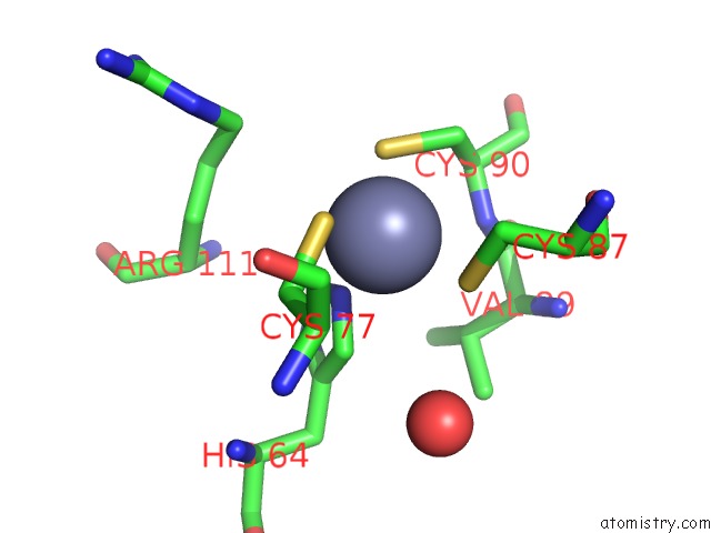
Zinc binding site 1 out of 2 in 2a1k
Go back to
Zinc binding site 1 out
of 2 in the RB69 Single-Stranded Dna Binding Protein Core Domain
Mono view

Stereo pair view
Mono view
Stereo pair view
A full contact list of Zinc with other atoms in the Zn binding
site number 1 of RB69 Single-Stranded Dna Binding Protein Core Domain within 5.0Å range:
|
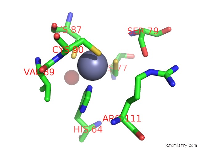
Zinc binding site 2 out of 2 in 2a1k
Go back to
Zinc binding site 2 out
of 2 in the RB69 Single-Stranded Dna Binding Protein Core Domain
Mono view

Stereo pair view
Mono view
Stereo pair view
A full contact list of Zinc with other atoms in the Zn binding
site number 2 of RB69 Single-Stranded Dna Binding Protein Core Domain within 5.0Å range:
|
Reference:
S.Sun,
L.Geng,
Y.Shamoo.
Structure and Enzymatic Properties of A Chimeric Bacteriophage RB69 Dna Polymerase and Single-Stranded Dna Binding Protein with Increased Processivity. Proteins V. 65 231 2006.
ISSN: ISSN 0887-3585
PubMed: 16881051
DOI: 10.1002/PROT.21088
Page generated: Wed Oct 16 21:28:24 2024
ISSN: ISSN 0887-3585
PubMed: 16881051
DOI: 10.1002/PROT.21088
Last articles
Zn in 9J0NZn in 9J0O
Zn in 9J0P
Zn in 9FJX
Zn in 9EKB
Zn in 9C0F
Zn in 9CAH
Zn in 9CH0
Zn in 9CH3
Zn in 9CH1