Zinc »
PDB 1z5r-1zfo »
1za1 »
Zinc in PDB 1za1: Structure of Wild-Type E. Coli Aspartate Transcarbamoylase in the Presence of Ctp at 2.20 A Resolution
Enzymatic activity of Structure of Wild-Type E. Coli Aspartate Transcarbamoylase in the Presence of Ctp at 2.20 A Resolution
All present enzymatic activity of Structure of Wild-Type E. Coli Aspartate Transcarbamoylase in the Presence of Ctp at 2.20 A Resolution:
2.1.3.2;
2.1.3.2;
Protein crystallography data
The structure of Structure of Wild-Type E. Coli Aspartate Transcarbamoylase in the Presence of Ctp at 2.20 A Resolution, PDB code: 1za1
was solved by
J.Wang,
K.A.Stieglitz,
J.P.Cardia,
E.R.Kantrowitz,
with X-Ray Crystallography technique. A brief refinement statistics is given in the table below:
| Resolution Low / High (Å) | 30.00 / 2.20 |
| Space group | P 3 2 1 |
| Cell size a, b, c (Å), α, β, γ (°) | 120.294, 120.294, 142.555, 90.00, 90.00, 120.00 |
| R / Rfree (%) | 20.5 / 25.2 |
Zinc Binding Sites:
The binding sites of Zinc atom in the Structure of Wild-Type E. Coli Aspartate Transcarbamoylase in the Presence of Ctp at 2.20 A Resolution
(pdb code 1za1). This binding sites where shown within
5.0 Angstroms radius around Zinc atom.
In total 2 binding sites of Zinc where determined in the Structure of Wild-Type E. Coli Aspartate Transcarbamoylase in the Presence of Ctp at 2.20 A Resolution, PDB code: 1za1:
Jump to Zinc binding site number: 1; 2;
In total 2 binding sites of Zinc where determined in the Structure of Wild-Type E. Coli Aspartate Transcarbamoylase in the Presence of Ctp at 2.20 A Resolution, PDB code: 1za1:
Jump to Zinc binding site number: 1; 2;
Zinc binding site 1 out of 2 in 1za1
Go back to
Zinc binding site 1 out
of 2 in the Structure of Wild-Type E. Coli Aspartate Transcarbamoylase in the Presence of Ctp at 2.20 A Resolution
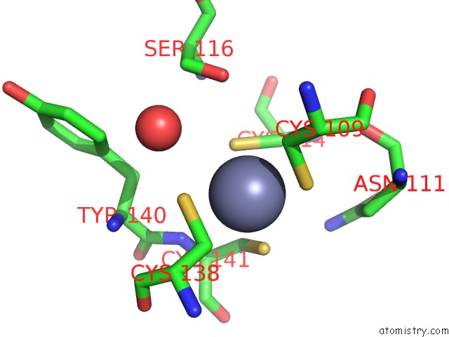
Mono view
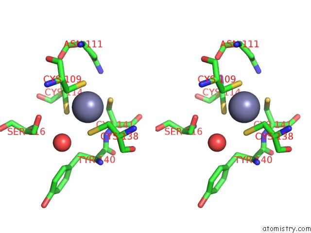
Stereo pair view
Mono view
Stereo pair view
A full contact list of Zinc with other atoms in the Zn binding
site number 1 of Structure of Wild-Type E. Coli Aspartate Transcarbamoylase in the Presence of Ctp at 2.20 A Resolution within 5.0Å range:
|
Zinc binding site 2 out of 2 in 1za1
Go back to
Zinc binding site 2 out
of 2 in the Structure of Wild-Type E. Coli Aspartate Transcarbamoylase in the Presence of Ctp at 2.20 A Resolution
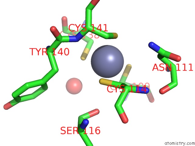
Mono view
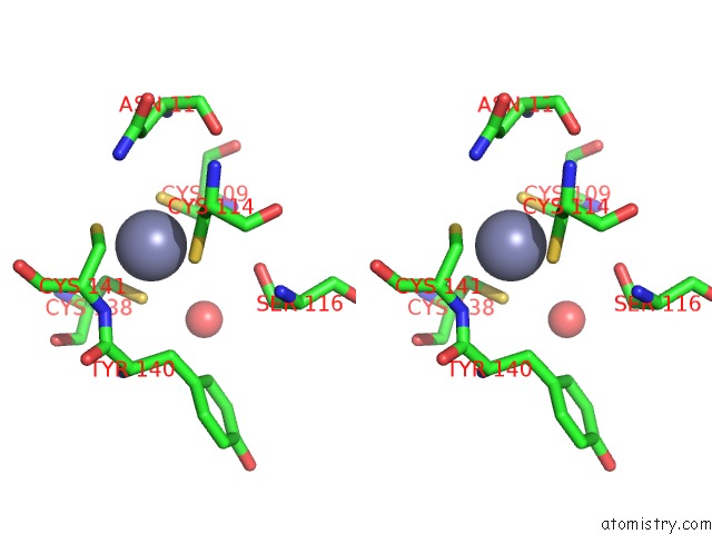
Stereo pair view
Mono view
Stereo pair view
A full contact list of Zinc with other atoms in the Zn binding
site number 2 of Structure of Wild-Type E. Coli Aspartate Transcarbamoylase in the Presence of Ctp at 2.20 A Resolution within 5.0Å range:
|
Reference:
J.Wang,
K.A.Stieglitz,
J.P.Cardia,
E.R.Kantrowitz.
Structural Basis For Ordered Substrate Binding and Cooperativity in Aspartate Transcarbamoylase Proc.Natl.Acad.Sci.Usa V. 102 8881 2005.
ISSN: ISSN 0027-8424
PubMed: 15951418
DOI: 10.1073/PNAS.0503742102
Page generated: Wed Oct 16 21:10:48 2024
ISSN: ISSN 0027-8424
PubMed: 15951418
DOI: 10.1073/PNAS.0503742102
Last articles
Zn in 9J0NZn in 9J0O
Zn in 9J0P
Zn in 9FJX
Zn in 9EKB
Zn in 9C0F
Zn in 9CAH
Zn in 9CH0
Zn in 9CH3
Zn in 9CH1