Zinc »
PDB 1z5r-1zfo »
1z9g »
Zinc in PDB 1z9g: Crystal Structure Analysis of Thermolysin Complexed with the Inhibitor (R)-Retro-Thiorphan
Enzymatic activity of Crystal Structure Analysis of Thermolysin Complexed with the Inhibitor (R)-Retro-Thiorphan
All present enzymatic activity of Crystal Structure Analysis of Thermolysin Complexed with the Inhibitor (R)-Retro-Thiorphan:
3.4.24.27;
3.4.24.27;
Protein crystallography data
The structure of Crystal Structure Analysis of Thermolysin Complexed with the Inhibitor (R)-Retro-Thiorphan, PDB code: 1z9g
was solved by
S.L.Roderick,
M.C.Fournie-Zaluski,
B.P.Roques,
B.W.Matthews,
with X-Ray Crystallography technique. A brief refinement statistics is given in the table below:
| Resolution Low / High (Å) | 10.00 / 1.70 |
| Space group | P 61 2 2 |
| Cell size a, b, c (Å), α, β, γ (°) | 94.000, 94.000, 132.000, 90.00, 90.00, 120.00 |
| R / Rfree (%) | n/a / n/a |
Other elements in 1z9g:
The structure of Crystal Structure Analysis of Thermolysin Complexed with the Inhibitor (R)-Retro-Thiorphan also contains other interesting chemical elements:
| Calcium | (Ca) | 4 atoms |
Zinc Binding Sites:
The binding sites of Zinc atom in the Crystal Structure Analysis of Thermolysin Complexed with the Inhibitor (R)-Retro-Thiorphan
(pdb code 1z9g). This binding sites where shown within
5.0 Angstroms radius around Zinc atom.
In total only one binding site of Zinc was determined in the Crystal Structure Analysis of Thermolysin Complexed with the Inhibitor (R)-Retro-Thiorphan, PDB code: 1z9g:
In total only one binding site of Zinc was determined in the Crystal Structure Analysis of Thermolysin Complexed with the Inhibitor (R)-Retro-Thiorphan, PDB code: 1z9g:
Zinc binding site 1 out of 1 in 1z9g
Go back to
Zinc binding site 1 out
of 1 in the Crystal Structure Analysis of Thermolysin Complexed with the Inhibitor (R)-Retro-Thiorphan
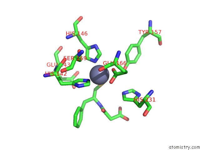
Mono view
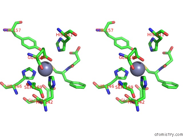
Stereo pair view
Mono view
Stereo pair view
A full contact list of Zinc with other atoms in the Zn binding
site number 1 of Crystal Structure Analysis of Thermolysin Complexed with the Inhibitor (R)-Retro-Thiorphan within 5.0Å range:
|
Reference:
S.L.Roderick,
M.C.Fournie-Zaluski,
B.P.Roques,
B.W.Matthews.
Thiorphan and Retro-Thiorphan Display Equivalent Interactions When Bound to Crystalline Thermolysin Biochemistry V. 28 1493 1989.
ISSN: ISSN 0006-2960
PubMed: 2719912
DOI: 10.1021/BI00430A011
Page generated: Wed Oct 16 21:09:56 2024
ISSN: ISSN 0006-2960
PubMed: 2719912
DOI: 10.1021/BI00430A011
Last articles
Zn in 9J0NZn in 9J0O
Zn in 9J0P
Zn in 9FJX
Zn in 9EKB
Zn in 9C0F
Zn in 9CAH
Zn in 9CH0
Zn in 9CH3
Zn in 9CH1