Zinc »
PDB 1x8h-1xm8 »
1xeg »
Zinc in PDB 1xeg: Crystal Structure of Human Carbonic Anhydrase II Complexed with An Acetate Ion
Enzymatic activity of Crystal Structure of Human Carbonic Anhydrase II Complexed with An Acetate Ion
All present enzymatic activity of Crystal Structure of Human Carbonic Anhydrase II Complexed with An Acetate Ion:
4.2.1.1;
4.2.1.1;
Protein crystallography data
The structure of Crystal Structure of Human Carbonic Anhydrase II Complexed with An Acetate Ion, PDB code: 1xeg
was solved by
P.A.Mazumdar,
D.Kumaran,
A.K.Das,
S.Swaminathan,
with X-Ray Crystallography technique. A brief refinement statistics is given in the table below:
| Resolution Low / High (Å) | 50.00 / 1.81 |
| Space group | P 21 21 21 |
| Cell size a, b, c (Å), α, β, γ (°) | 42.312, 71.749, 74.010, 90.00, 90.00, 90.00 |
| R / Rfree (%) | 18 / 21 |
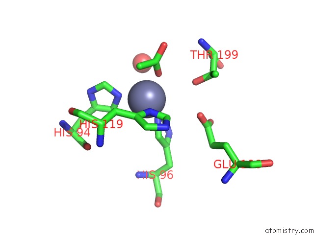
Zinc Binding Sites:
The binding sites of Zinc atom in the Crystal Structure of Human Carbonic Anhydrase II Complexed with An Acetate Ion
(pdb code 1xeg). This binding sites where shown within
5.0 Angstroms radius around Zinc atom.
In total only one binding site of Zinc was determined in the Crystal Structure of Human Carbonic Anhydrase II Complexed with An Acetate Ion, PDB code: 1xeg:
In total only one binding site of Zinc was determined in the Crystal Structure of Human Carbonic Anhydrase II Complexed with An Acetate Ion, PDB code: 1xeg:
Zinc binding site 1 out of 1 in 1xeg
Go back to
Zinc binding site 1 out
of 1 in the Crystal Structure of Human Carbonic Anhydrase II Complexed with An Acetate Ion
Mono view

Stereo pair view
Mono view
Stereo pair view
A full contact list of Zinc with other atoms in the Zn binding
site number 1 of Crystal Structure of Human Carbonic Anhydrase II Complexed with An Acetate Ion within 5.0Å range:
|
Reference:
P.A.Mazumdar,
D.Kumaran,
S.Swaminathan,
A.K.Das.
A Novel Acetate-Bound Complex of Human Carbonic Anhydrase II. Acta Crystallogr.,Sect.F V. 64 163 2008.
ISSN: ESSN 1744-3091
PubMed: 18323598
DOI: 10.1107/S1744309108002078
Page generated: Wed Aug 20 00:17:20 2025
ISSN: ESSN 1744-3091
PubMed: 18323598
DOI: 10.1107/S1744309108002078
Last articles
Zn in 2GTQZn in 2GU1
Zn in 2GTL
Zn in 2GSU
Zn in 2GSN
Zn in 2GSO
Zn in 2GPY
Zn in 2GQJ
Zn in 2GP5
Zn in 2GQE