Zinc »
PDB 1w5m-1wfx »
1w8p »
Zinc in PDB 1w8p: Structural Properties of the B25TYR-Nme-B26PHE Insulin Mutant.
Protein crystallography data
The structure of Structural Properties of the B25TYR-Nme-B26PHE Insulin Mutant., PDB code: 1w8p
was solved by
L.Zakowa,
O.Au-Alvarez,
E.J.Dodson,
G.G.Dodson,
A.M.Brzozowski,
with X-Ray Crystallography technique. A brief refinement statistics is given in the table below:
| Resolution Low / High (Å) | 25.57 / 2.08 |
| Space group | P 1 21 1 |
| Cell size a, b, c (Å), α, β, γ (°) | 59.903, 62.116, 47.796, 90.00, 110.58, 90.00 |
| R / Rfree (%) | 18.9 / 25.6 |
Zinc Binding Sites:
The binding sites of Zinc atom in the Structural Properties of the B25TYR-Nme-B26PHE Insulin Mutant.
(pdb code 1w8p). This binding sites where shown within
5.0 Angstroms radius around Zinc atom.
In total 2 binding sites of Zinc where determined in the Structural Properties of the B25TYR-Nme-B26PHE Insulin Mutant., PDB code: 1w8p:
Jump to Zinc binding site number: 1; 2;
In total 2 binding sites of Zinc where determined in the Structural Properties of the B25TYR-Nme-B26PHE Insulin Mutant., PDB code: 1w8p:
Jump to Zinc binding site number: 1; 2;
Zinc binding site 1 out of 2 in 1w8p
Go back to
Zinc binding site 1 out
of 2 in the Structural Properties of the B25TYR-Nme-B26PHE Insulin Mutant.
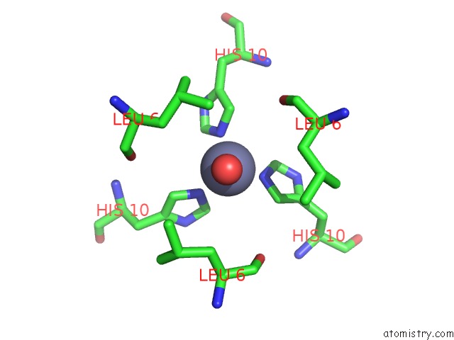
Mono view
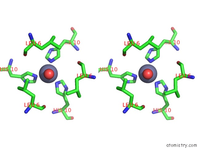
Stereo pair view
Mono view
Stereo pair view
A full contact list of Zinc with other atoms in the Zn binding
site number 1 of Structural Properties of the B25TYR-Nme-B26PHE Insulin Mutant. within 5.0Å range:
|
Zinc binding site 2 out of 2 in 1w8p
Go back to
Zinc binding site 2 out
of 2 in the Structural Properties of the B25TYR-Nme-B26PHE Insulin Mutant.
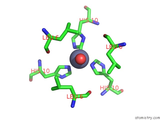
Mono view
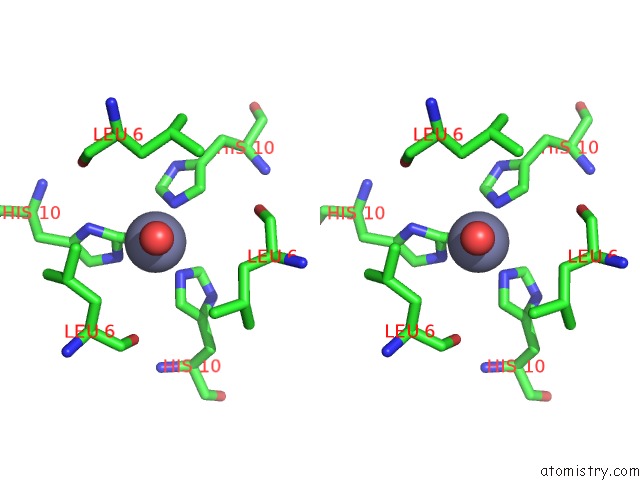
Stereo pair view
Mono view
Stereo pair view
A full contact list of Zinc with other atoms in the Zn binding
site number 2 of Structural Properties of the B25TYR-Nme-B26PHE Insulin Mutant. within 5.0Å range:
|
Reference:
L.Zarkowa,
J.Brynda,
O.Au-Alvarez,
E.J.Dodson,
G.G.Dodson,
J.L.Whittingham,
A.M.Brzozowski.
Towards the Insulin-Igf-I Intermediate Structures: Functional and Structural Properties of the B25TYR-Nme-B26PHE Insulin Mutant. Biochemistry V. 43 16293 2004.
ISSN: ISSN 0006-2960
PubMed: 15610023
DOI: 10.1021/BI048856U
Page generated: Wed Oct 16 19:56:21 2024
ISSN: ISSN 0006-2960
PubMed: 15610023
DOI: 10.1021/BI048856U
Last articles
Zn in 9J0NZn in 9J0O
Zn in 9J0P
Zn in 9FJX
Zn in 9EKB
Zn in 9C0F
Zn in 9CAH
Zn in 9CH0
Zn in 9CH3
Zn in 9CH1