Zinc »
PDB 1ul4-1v13 »
1urj »
Zinc in PDB 1urj: Single Stranded Dna-Binding Protein(ICP8) From Herpes Simplex Virus-1
Protein crystallography data
The structure of Single Stranded Dna-Binding Protein(ICP8) From Herpes Simplex Virus-1, PDB code: 1urj
was solved by
S.Panjikar,
M.Mapelli,
P.A.Tucker,
with X-Ray Crystallography technique. A brief refinement statistics is given in the table below:
| Resolution Low / High (Å) | 20.00 / 3.00 |
| Space group | P 21 21 21 |
| Cell size a, b, c (Å), α, β, γ (°) | 100.910, 145.370, 162.030, 90.00, 90.00, 90.00 |
| R / Rfree (%) | 23.5 / 28.6 |
Other elements in 1urj:
The structure of Single Stranded Dna-Binding Protein(ICP8) From Herpes Simplex Virus-1 also contains other interesting chemical elements:
| Mercury | (Hg) | 8 atoms |
Zinc Binding Sites:
The binding sites of Zinc atom in the Single Stranded Dna-Binding Protein(ICP8) From Herpes Simplex Virus-1
(pdb code 1urj). This binding sites where shown within
5.0 Angstroms radius around Zinc atom.
In total 2 binding sites of Zinc where determined in the Single Stranded Dna-Binding Protein(ICP8) From Herpes Simplex Virus-1, PDB code: 1urj:
Jump to Zinc binding site number: 1; 2;
In total 2 binding sites of Zinc where determined in the Single Stranded Dna-Binding Protein(ICP8) From Herpes Simplex Virus-1, PDB code: 1urj:
Jump to Zinc binding site number: 1; 2;
Zinc binding site 1 out of 2 in 1urj
Go back to
Zinc binding site 1 out
of 2 in the Single Stranded Dna-Binding Protein(ICP8) From Herpes Simplex Virus-1
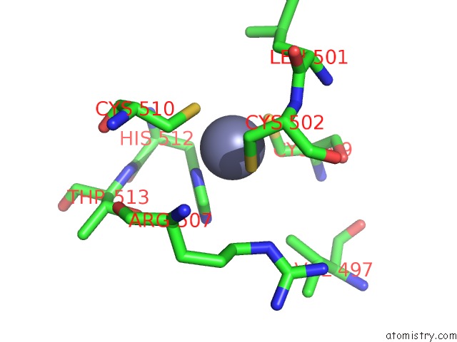
Mono view
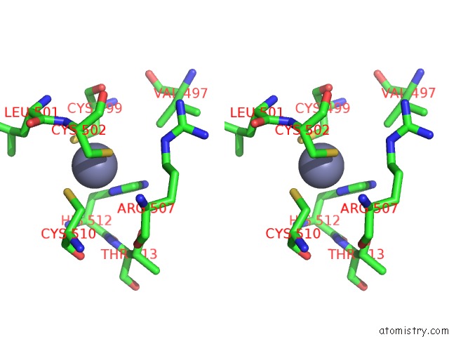
Stereo pair view
Mono view
Stereo pair view
A full contact list of Zinc with other atoms in the Zn binding
site number 1 of Single Stranded Dna-Binding Protein(ICP8) From Herpes Simplex Virus-1 within 5.0Å range:
|
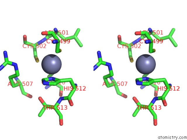
Zinc binding site 2 out of 2 in 1urj
Go back to
Zinc binding site 2 out
of 2 in the Single Stranded Dna-Binding Protein(ICP8) From Herpes Simplex Virus-1
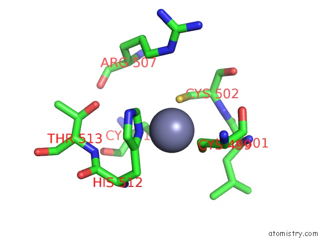
Mono view
Stereo pair view
Mono view
Stereo pair view
A full contact list of Zinc with other atoms in the Zn binding
site number 2 of Single Stranded Dna-Binding Protein(ICP8) From Herpes Simplex Virus-1 within 5.0Å range:
|
Reference:
M.Mapelli,
S.Panjikar,
P.A.Tucker.
The Crystal Structure of the Herpes Simplex Virus 1 Ssdna-Binding Protein Suggests the Structural Basis For Flexible, Cooperative Single-Stranded Dna Binding. J. Biol. Chem. V. 280 2990 2005.
ISSN: ISSN 0021-9258
PubMed: 15507432
DOI: 10.1074/JBC.M406780200
Page generated: Wed Oct 16 19:36:06 2024
ISSN: ISSN 0021-9258
PubMed: 15507432
DOI: 10.1074/JBC.M406780200
Last articles
Zn in 9J0NZn in 9J0O
Zn in 9J0P
Zn in 9FJX
Zn in 9EKB
Zn in 9C0F
Zn in 9CAH
Zn in 9CH0
Zn in 9CH3
Zn in 9CH1