Zinc »
PDB 1ul4-1v13 »
1un6 »
Zinc in PDB 1un6: The Crystal Structure of A Zinc Finger - Rna Complex Reveals Two Modes of Molecular Recognition
Protein crystallography data
The structure of The Crystal Structure of A Zinc Finger - Rna Complex Reveals Two Modes of Molecular Recognition, PDB code: 1un6
was solved by
D.Lu,
M.A.Searles,
A.Klug,
with X-Ray Crystallography technique. A brief refinement statistics is given in the table below:
| Resolution Low / High (Å) | 35.19 / 3.10 |
| Space group | C 1 2 1 |
| Cell size a, b, c (Å), α, β, γ (°) | 58.598, 191.593, 79.770, 90.00, 101.51, 90.00 |
| R / Rfree (%) | 21.6 / 25.9 |
Other elements in 1un6:
The structure of The Crystal Structure of A Zinc Finger - Rna Complex Reveals Two Modes of Molecular Recognition also contains other interesting chemical elements:
| Magnesium | (Mg) | 13 atoms |
Zinc Binding Sites:
The binding sites of Zinc atom in the The Crystal Structure of A Zinc Finger - Rna Complex Reveals Two Modes of Molecular Recognition
(pdb code 1un6). This binding sites where shown within
5.0 Angstroms radius around Zinc atom.
In total 8 binding sites of Zinc where determined in the The Crystal Structure of A Zinc Finger - Rna Complex Reveals Two Modes of Molecular Recognition, PDB code: 1un6:
Jump to Zinc binding site number: 1; 2; 3; 4; 5; 6; 7; 8;
In total 8 binding sites of Zinc where determined in the The Crystal Structure of A Zinc Finger - Rna Complex Reveals Two Modes of Molecular Recognition, PDB code: 1un6:
Jump to Zinc binding site number: 1; 2; 3; 4; 5; 6; 7; 8;
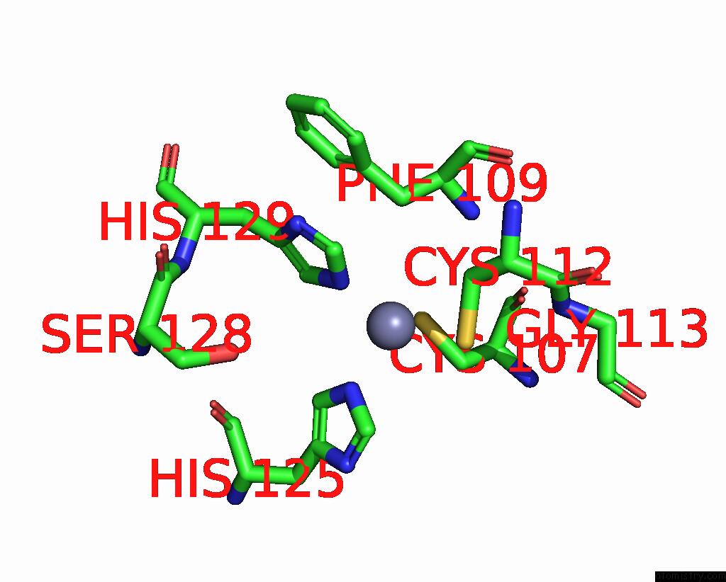
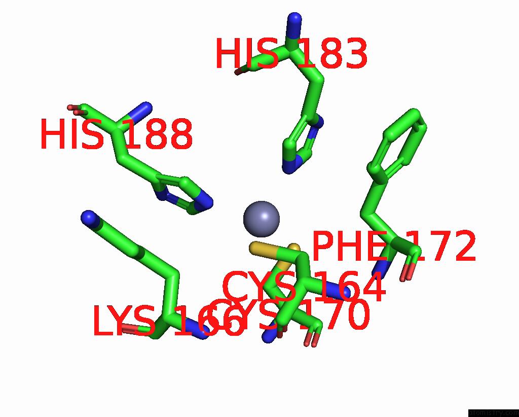
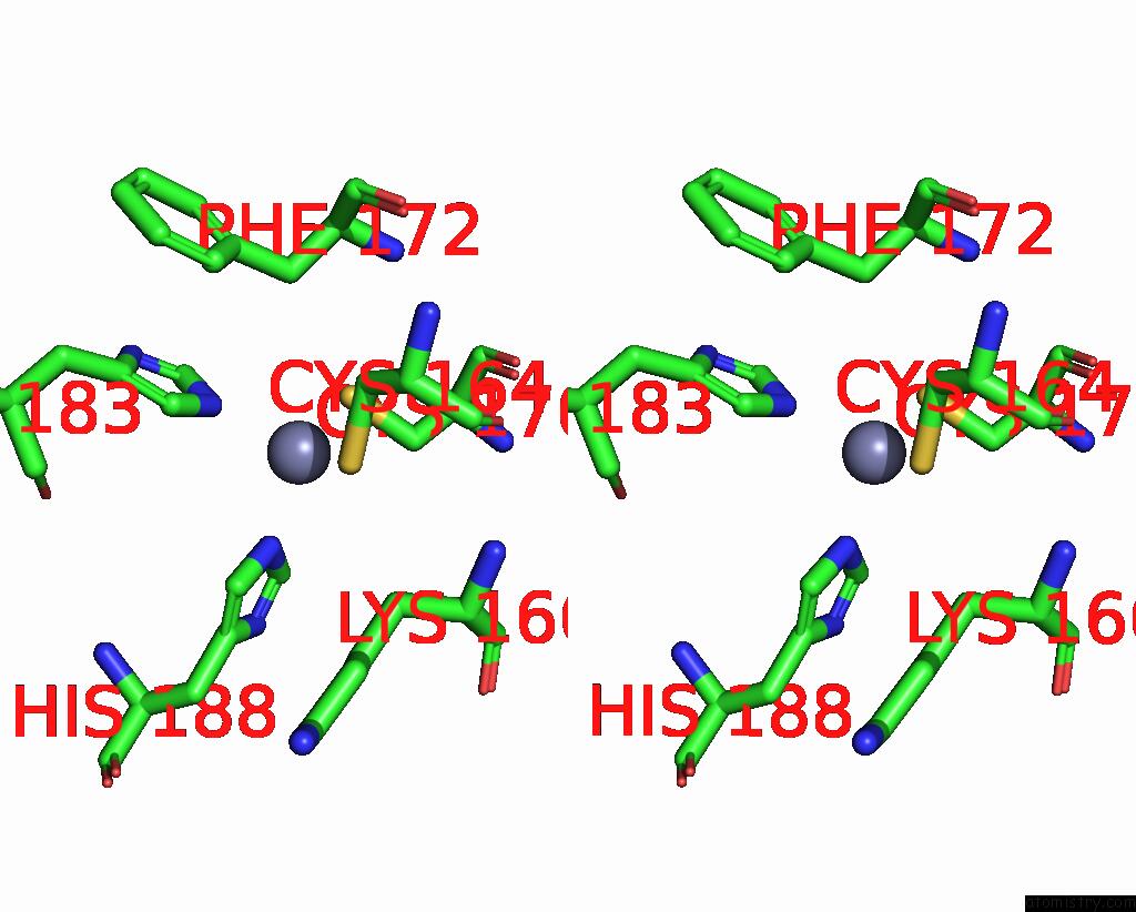
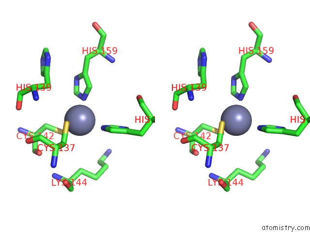
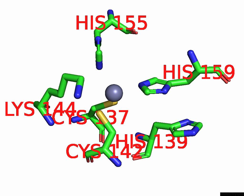
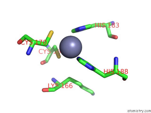
Zinc binding site 1 out of 8 in 1un6
Go back to
Zinc binding site 1 out
of 8 in the The Crystal Structure of A Zinc Finger - Rna Complex Reveals Two Modes of Molecular Recognition
Mono view
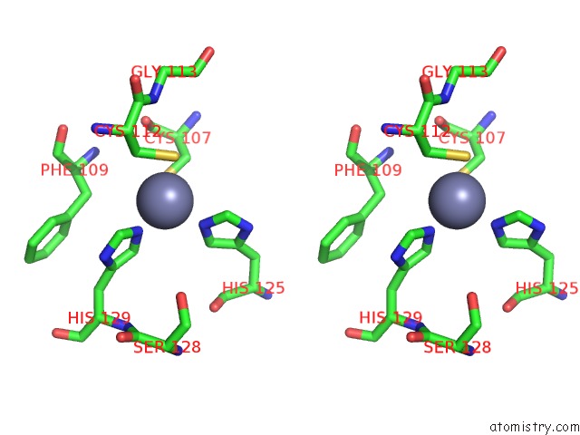
Stereo pair view
Mono view
Stereo pair view
A full contact list of Zinc with other atoms in the Zn binding
site number 1 of The Crystal Structure of A Zinc Finger - Rna Complex Reveals Two Modes of Molecular Recognition within 5.0Å range:
|
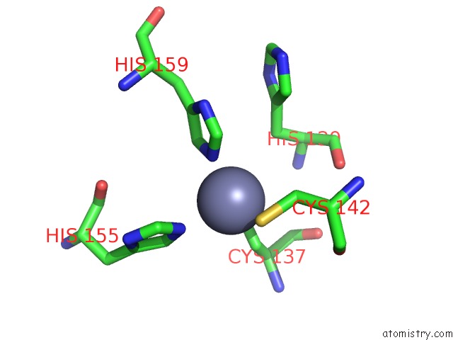
Zinc binding site 2 out of 8 in 1un6
Go back to
Zinc binding site 2 out
of 8 in the The Crystal Structure of A Zinc Finger - Rna Complex Reveals Two Modes of Molecular Recognition
Mono view
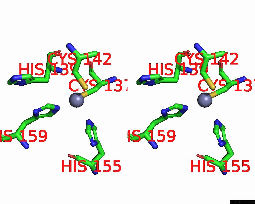
Stereo pair view
Mono view
Stereo pair view
A full contact list of Zinc with other atoms in the Zn binding
site number 2 of The Crystal Structure of A Zinc Finger - Rna Complex Reveals Two Modes of Molecular Recognition within 5.0Å range:
|
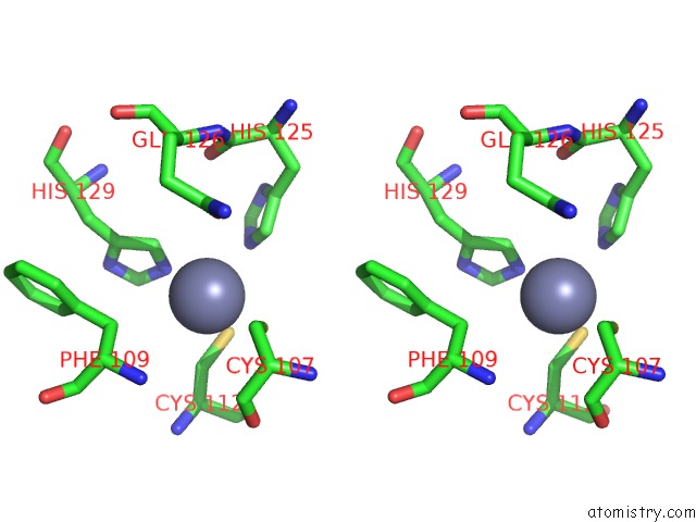
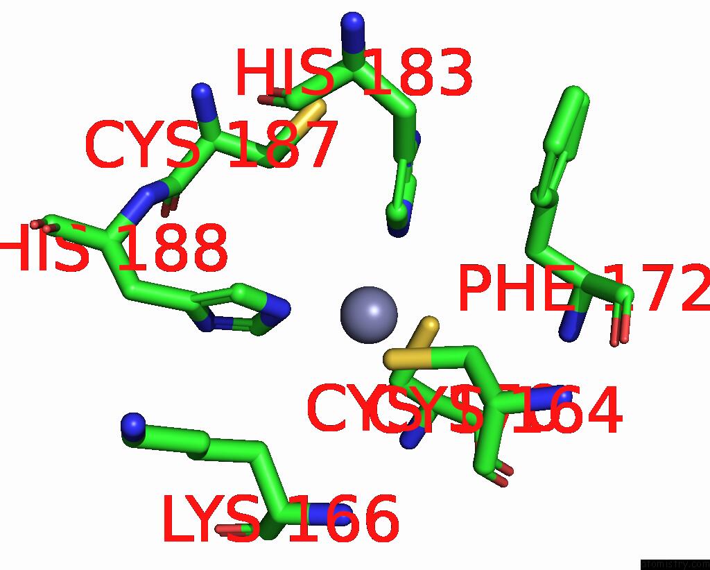
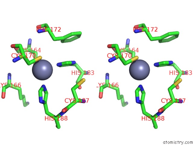
Zinc binding site 3 out of 8 in 1un6
Go back to
Zinc binding site 3 out
of 8 in the The Crystal Structure of A Zinc Finger - Rna Complex Reveals Two Modes of Molecular Recognition
Mono view
Stereo pair view
Mono view
Stereo pair view
A full contact list of Zinc with other atoms in the Zn binding
site number 3 of The Crystal Structure of A Zinc Finger - Rna Complex Reveals Two Modes of Molecular Recognition within 5.0Å range:
|
Zinc binding site 4 out of 8 in 1un6
Go back to
Zinc binding site 4 out
of 8 in the The Crystal Structure of A Zinc Finger - Rna Complex Reveals Two Modes of Molecular Recognition

Mono view
Stereo pair view
Mono view
Stereo pair view
A full contact list of Zinc with other atoms in the Zn binding
site number 4 of The Crystal Structure of A Zinc Finger - Rna Complex Reveals Two Modes of Molecular Recognition within 5.0Å range:
|
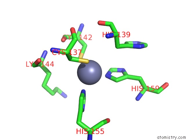
Zinc binding site 5 out of 8 in 1un6
Go back to
Zinc binding site 5 out
of 8 in the The Crystal Structure of A Zinc Finger - Rna Complex Reveals Two Modes of Molecular Recognition
Mono view
Stereo pair view
Mono view
Stereo pair view
A full contact list of Zinc with other atoms in the Zn binding
site number 5 of The Crystal Structure of A Zinc Finger - Rna Complex Reveals Two Modes of Molecular Recognition within 5.0Å range:
|
Zinc binding site 6 out of 8 in 1un6
Go back to
Zinc binding site 6 out
of 8 in the The Crystal Structure of A Zinc Finger - Rna Complex Reveals Two Modes of Molecular Recognition
Mono view
Stereo pair view
Mono view
Stereo pair view
A full contact list of Zinc with other atoms in the Zn binding
site number 6 of The Crystal Structure of A Zinc Finger - Rna Complex Reveals Two Modes of Molecular Recognition within 5.0Å range:
|
Zinc binding site 7 out of 8 in 1un6
Go back to
Zinc binding site 7 out
of 8 in the The Crystal Structure of A Zinc Finger - Rna Complex Reveals Two Modes of Molecular Recognition
Mono view

Stereo pair view
Mono view
Stereo pair view
A full contact list of Zinc with other atoms in the Zn binding
site number 7 of The Crystal Structure of A Zinc Finger - Rna Complex Reveals Two Modes of Molecular Recognition within 5.0Å range:
|
Zinc binding site 8 out of 8 in 1un6
Go back to
Zinc binding site 8 out
of 8 in the The Crystal Structure of A Zinc Finger - Rna Complex Reveals Two Modes of Molecular Recognition
Mono view

Stereo pair view
Mono view
Stereo pair view
A full contact list of Zinc with other atoms in the Zn binding
site number 8 of The Crystal Structure of A Zinc Finger - Rna Complex Reveals Two Modes of Molecular Recognition within 5.0Å range:
|
Reference:
D.Lu,
M.A.Searles,
A.Klug.
Crystal Structure of A Zinc-Finger-Rna Complex Reveals Two Modes of Molecular Recognition Nature V. 426 96 2003.
ISSN: ISSN 0028-0836
PubMed: 14603324
DOI: 10.1038/NATURE02088
Page generated: Wed Oct 16 19:33:56 2024
ISSN: ISSN 0028-0836
PubMed: 14603324
DOI: 10.1038/NATURE02088
Last articles
Zn in 9J0NZn in 9J0O
Zn in 9J0P
Zn in 9FJX
Zn in 9EKB
Zn in 9C0F
Zn in 9CAH
Zn in 9CH0
Zn in 9CH3
Zn in 9CH1