Zinc »
PDB 1ul4-1v13 »
1umt »
Zinc in PDB 1umt: Stromelysin-1 Catalytic Domain with Hydrophobic Inhibitor Bound, pH 7.0, 32OC, 20 Mm CACL2, 15% Acetonitrile; uc(Nmr) Average of 20 Structures Minimized with Restraints
Enzymatic activity of Stromelysin-1 Catalytic Domain with Hydrophobic Inhibitor Bound, pH 7.0, 32OC, 20 Mm CACL2, 15% Acetonitrile; uc(Nmr) Average of 20 Structures Minimized with Restraints
All present enzymatic activity of Stromelysin-1 Catalytic Domain with Hydrophobic Inhibitor Bound, pH 7.0, 32OC, 20 Mm CACL2, 15% Acetonitrile; uc(Nmr) Average of 20 Structures Minimized with Restraints:
3.4.24.17;
3.4.24.17;
Other elements in 1umt:
The structure of Stromelysin-1 Catalytic Domain with Hydrophobic Inhibitor Bound, pH 7.0, 32OC, 20 Mm CACL2, 15% Acetonitrile; uc(Nmr) Average of 20 Structures Minimized with Restraints also contains other interesting chemical elements:
| Calcium | (Ca) | 1 atom |
Zinc Binding Sites:
The binding sites of Zinc atom in the Stromelysin-1 Catalytic Domain with Hydrophobic Inhibitor Bound, pH 7.0, 32OC, 20 Mm CACL2, 15% Acetonitrile; uc(Nmr) Average of 20 Structures Minimized with Restraints
(pdb code 1umt). This binding sites where shown within
5.0 Angstroms radius around Zinc atom.
In total 2 binding sites of Zinc where determined in the Stromelysin-1 Catalytic Domain with Hydrophobic Inhibitor Bound, pH 7.0, 32OC, 20 Mm CACL2, 15% Acetonitrile; uc(Nmr) Average of 20 Structures Minimized with Restraints, PDB code: 1umt:
Jump to Zinc binding site number: 1; 2;
In total 2 binding sites of Zinc where determined in the Stromelysin-1 Catalytic Domain with Hydrophobic Inhibitor Bound, pH 7.0, 32OC, 20 Mm CACL2, 15% Acetonitrile; uc(Nmr) Average of 20 Structures Minimized with Restraints, PDB code: 1umt:
Jump to Zinc binding site number: 1; 2;
Zinc binding site 1 out of 2 in 1umt
Go back to
Zinc binding site 1 out
of 2 in the Stromelysin-1 Catalytic Domain with Hydrophobic Inhibitor Bound, pH 7.0, 32OC, 20 Mm CACL2, 15% Acetonitrile; uc(Nmr) Average of 20 Structures Minimized with Restraints
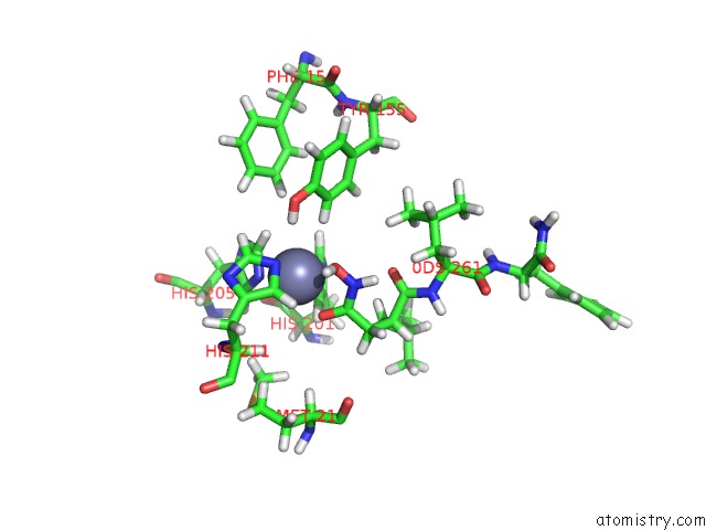
Mono view
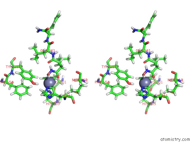
Stereo pair view
Mono view
Stereo pair view
A full contact list of Zinc with other atoms in the Zn binding
site number 1 of Stromelysin-1 Catalytic Domain with Hydrophobic Inhibitor Bound, pH 7.0, 32OC, 20 Mm CACL2, 15% Acetonitrile; uc(Nmr) Average of 20 Structures Minimized with Restraints within 5.0Å range:
|
Zinc binding site 2 out of 2 in 1umt
Go back to
Zinc binding site 2 out
of 2 in the Stromelysin-1 Catalytic Domain with Hydrophobic Inhibitor Bound, pH 7.0, 32OC, 20 Mm CACL2, 15% Acetonitrile; uc(Nmr) Average of 20 Structures Minimized with Restraints
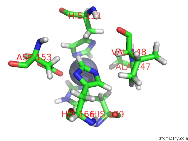
Mono view
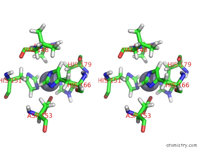
Stereo pair view
Mono view
Stereo pair view
A full contact list of Zinc with other atoms in the Zn binding
site number 2 of Stromelysin-1 Catalytic Domain with Hydrophobic Inhibitor Bound, pH 7.0, 32OC, 20 Mm CACL2, 15% Acetonitrile; uc(Nmr) Average of 20 Structures Minimized with Restraints within 5.0Å range:
|
Reference:
S.R.Van Doren,
A.V.Kurochkin,
W.Hu,
Q.Z.Ye,
L.L.Johnson,
D.J.Hupe,
E.R.Zuiderweg.
Solution Structure of the Catalytic Domain of Human Stromelysin Complexed with A Hydrophobic Inhibitor. Protein Sci. V. 4 2487 1995.
ISSN: ISSN 0961-8368
PubMed: 8580839
Page generated: Wed Oct 16 19:33:52 2024
ISSN: ISSN 0961-8368
PubMed: 8580839
Last articles
Zn in 9J0NZn in 9J0O
Zn in 9J0P
Zn in 9FJX
Zn in 9EKB
Zn in 9C0F
Zn in 9CAH
Zn in 9CH0
Zn in 9CH3
Zn in 9CH1