Zinc »
PDB 1u2n-1uip »
1ugc »
Zinc in PDB 1ugc: Human Carbonic Anhydrase II [Hcaii] (E.C.4.2.1.1) Mutant with Ala 65 Replaced By His (A65H)
Enzymatic activity of Human Carbonic Anhydrase II [Hcaii] (E.C.4.2.1.1) Mutant with Ala 65 Replaced By His (A65H)
All present enzymatic activity of Human Carbonic Anhydrase II [Hcaii] (E.C.4.2.1.1) Mutant with Ala 65 Replaced By His (A65H):
4.2.1.1;
4.2.1.1;
Protein crystallography data
The structure of Human Carbonic Anhydrase II [Hcaii] (E.C.4.2.1.1) Mutant with Ala 65 Replaced By His (A65H), PDB code: 1ugc
was solved by
L.R.Scolnick,
D.W.Christianson,
with X-Ray Crystallography technique. A brief refinement statistics is given in the table below:
| Resolution Low / High (Å) | 6.50 / 2.00 |
| Space group | P 1 21 1 |
| Cell size a, b, c (Å), α, β, γ (°) | 42.700, 41.700, 73.000, 90.00, 104.60, 90.00 |
| R / Rfree (%) | 16.2 / 22.5 |
Other elements in 1ugc:
The structure of Human Carbonic Anhydrase II [Hcaii] (E.C.4.2.1.1) Mutant with Ala 65 Replaced By His (A65H) also contains other interesting chemical elements:
| Mercury | (Hg) | 1 atom |
Zinc Binding Sites:
The binding sites of Zinc atom in the Human Carbonic Anhydrase II [Hcaii] (E.C.4.2.1.1) Mutant with Ala 65 Replaced By His (A65H)
(pdb code 1ugc). This binding sites where shown within
5.0 Angstroms radius around Zinc atom.
In total only one binding site of Zinc was determined in the Human Carbonic Anhydrase II [Hcaii] (E.C.4.2.1.1) Mutant with Ala 65 Replaced By His (A65H), PDB code: 1ugc:
In total only one binding site of Zinc was determined in the Human Carbonic Anhydrase II [Hcaii] (E.C.4.2.1.1) Mutant with Ala 65 Replaced By His (A65H), PDB code: 1ugc:
Zinc binding site 1 out of 1 in 1ugc
Go back to
Zinc binding site 1 out
of 1 in the Human Carbonic Anhydrase II [Hcaii] (E.C.4.2.1.1) Mutant with Ala 65 Replaced By His (A65H)
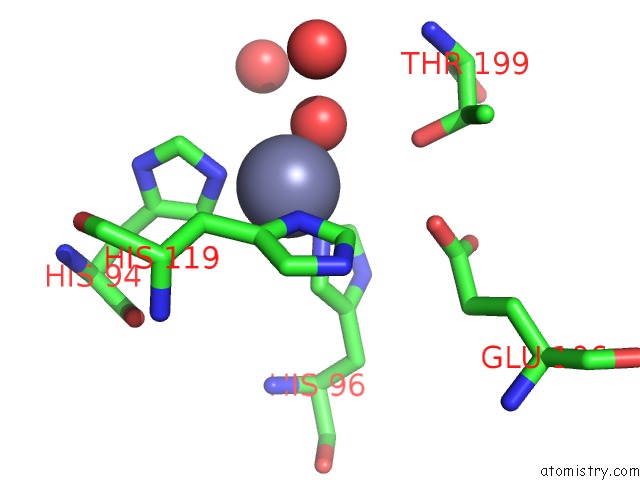
Mono view
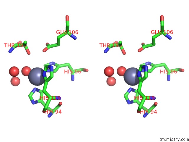
Stereo pair view
Mono view
Stereo pair view
A full contact list of Zinc with other atoms in the Zn binding
site number 1 of Human Carbonic Anhydrase II [Hcaii] (E.C.4.2.1.1) Mutant with Ala 65 Replaced By His (A65H) within 5.0Å range:
|
Reference:
L.R.Scolnick,
D.W.Christianson.
X-Ray Crystallographic Studies of Alanine-65 Variants of Carbonic Anhydrase II Reveal the Structural Basis of Compromised Proton Transfer in Catalysis. Biochemistry V. 35 16429 1996.
ISSN: ISSN 0006-2960
PubMed: 8987974
DOI: 10.1021/BI9617872
Page generated: Wed Oct 16 19:31:04 2024
ISSN: ISSN 0006-2960
PubMed: 8987974
DOI: 10.1021/BI9617872
Last articles
Zn in 9J0NZn in 9J0O
Zn in 9J0P
Zn in 9FJX
Zn in 9EKB
Zn in 9C0F
Zn in 9CAH
Zn in 9CH0
Zn in 9CH3
Zn in 9CH1