Zinc »
PDB 1to5-1u22 »
1tqx »
Zinc in PDB 1tqx: Crystal Structure of PFAL009167 A Putative D-Ribulose 5-Phosphate 3- Epimerase From P.Falciparum
Protein crystallography data
The structure of Crystal Structure of PFAL009167 A Putative D-Ribulose 5-Phosphate 3- Epimerase From P.Falciparum, PDB code: 1tqx
was solved by
J.Caruthers,
J.Bosch,
W.G.J.Hol,
Structural Genomics Of Pathogenicprotozoa Consortium (Sgpp),
with X-Ray Crystallography technique. A brief refinement statistics is given in the table below:
| Resolution Low / High (Å) | 52.17 / 2.00 |
| Space group | P 21 21 21 |
| Cell size a, b, c (Å), α, β, γ (°) | 79.415, 84.454, 120.717, 90.00, 90.00, 90.00 |
| R / Rfree (%) | 21.2 / 23.3 |
Zinc Binding Sites:
The binding sites of Zinc atom in the Crystal Structure of PFAL009167 A Putative D-Ribulose 5-Phosphate 3- Epimerase From P.Falciparum
(pdb code 1tqx). This binding sites where shown within
5.0 Angstroms radius around Zinc atom.
In total 2 binding sites of Zinc where determined in the Crystal Structure of PFAL009167 A Putative D-Ribulose 5-Phosphate 3- Epimerase From P.Falciparum, PDB code: 1tqx:
Jump to Zinc binding site number: 1; 2;
In total 2 binding sites of Zinc where determined in the Crystal Structure of PFAL009167 A Putative D-Ribulose 5-Phosphate 3- Epimerase From P.Falciparum, PDB code: 1tqx:
Jump to Zinc binding site number: 1; 2;
Zinc binding site 1 out of 2 in 1tqx
Go back to
Zinc binding site 1 out
of 2 in the Crystal Structure of PFAL009167 A Putative D-Ribulose 5-Phosphate 3- Epimerase From P.Falciparum
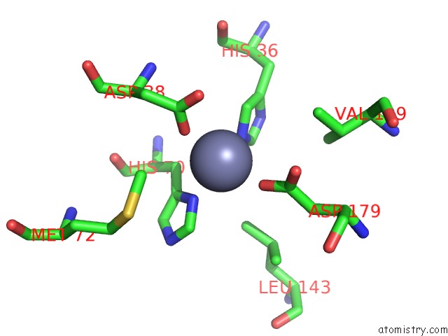
Mono view
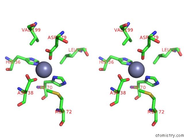
Stereo pair view
Mono view
Stereo pair view
A full contact list of Zinc with other atoms in the Zn binding
site number 1 of Crystal Structure of PFAL009167 A Putative D-Ribulose 5-Phosphate 3- Epimerase From P.Falciparum within 5.0Å range:
|
Zinc binding site 2 out of 2 in 1tqx
Go back to
Zinc binding site 2 out
of 2 in the Crystal Structure of PFAL009167 A Putative D-Ribulose 5-Phosphate 3- Epimerase From P.Falciparum
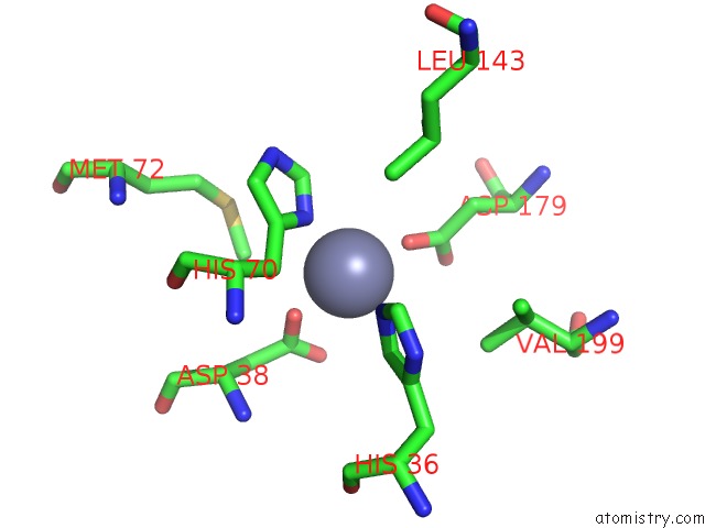
Mono view

Stereo pair view
Mono view
Stereo pair view
A full contact list of Zinc with other atoms in the Zn binding
site number 2 of Crystal Structure of PFAL009167 A Putative D-Ribulose 5-Phosphate 3- Epimerase From P.Falciparum within 5.0Å range:
|
Reference:
J.Caruthers,
J.Bosch,
F.Buckner,
W.Van Voorhis,
P.Myler,
E.Worthey,
C.Mehlin,
E.Boni,
G.Detitta,
J.Luft,
A.Lauricella,
O.Kalyuzhniy,
L.Anderson,
F.Zucker,
M.Soltis,
W.G.Hol.
Structure of A Ribulose 5-Phosphate 3-Epimerase From Plasmodium Falciparum. Proteins V. 62 338 2006.
ISSN: ISSN 0887-3585
PubMed: 16304640
DOI: 10.1002/PROT.20764
Page generated: Wed Oct 16 19:16:48 2024
ISSN: ISSN 0887-3585
PubMed: 16304640
DOI: 10.1002/PROT.20764
Last articles
Zn in 9J0NZn in 9J0O
Zn in 9J0P
Zn in 9FJX
Zn in 9EKB
Zn in 9C0F
Zn in 9CAH
Zn in 9CH0
Zn in 9CH3
Zn in 9CH1