Zinc »
PDB 1tbt-1to4 »
1thj »
Zinc in PDB 1thj: Carbonic Anhydrase From Methanosarcina
Enzymatic activity of Carbonic Anhydrase From Methanosarcina
All present enzymatic activity of Carbonic Anhydrase From Methanosarcina:
4.2.1.1;
4.2.1.1;
Protein crystallography data
The structure of Carbonic Anhydrase From Methanosarcina, PDB code: 1thj
was solved by
C.Kisker,
H.Schindelin,
D.C.Rees,
with X-Ray Crystallography technique. A brief refinement statistics is given in the table below:
| Resolution Low / High (Å) | 10.00 / 2.80 |
| Space group | P 43 21 2 |
| Cell size a, b, c (Å), α, β, γ (°) | 71.840, 71.840, 333.513, 90.00, 90.00, 90.00 |
| R / Rfree (%) | 23.4 / 29.5 |
Zinc Binding Sites:
The binding sites of Zinc atom in the Carbonic Anhydrase From Methanosarcina
(pdb code 1thj). This binding sites where shown within
5.0 Angstroms radius around Zinc atom.
In total 3 binding sites of Zinc where determined in the Carbonic Anhydrase From Methanosarcina, PDB code: 1thj:
Jump to Zinc binding site number: 1; 2; 3;
In total 3 binding sites of Zinc where determined in the Carbonic Anhydrase From Methanosarcina, PDB code: 1thj:
Jump to Zinc binding site number: 1; 2; 3;
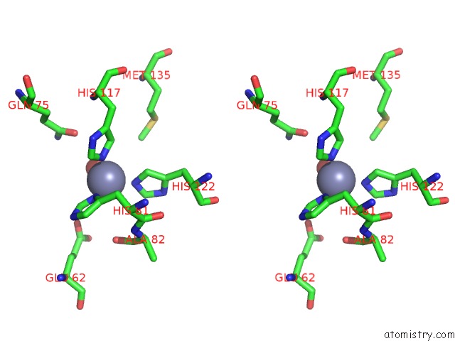
Zinc binding site 1 out of 3 in 1thj
Go back to
Zinc binding site 1 out
of 3 in the Carbonic Anhydrase From Methanosarcina
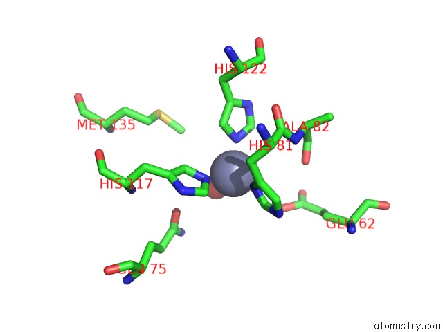
Mono view
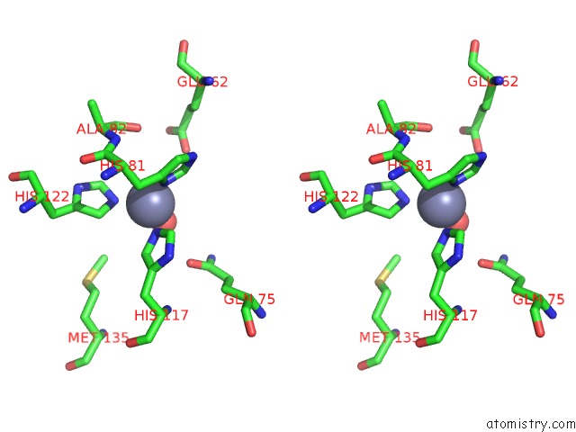
Stereo pair view
Mono view
Stereo pair view
A full contact list of Zinc with other atoms in the Zn binding
site number 1 of Carbonic Anhydrase From Methanosarcina within 5.0Å range:
|
Zinc binding site 2 out of 3 in 1thj
Go back to
Zinc binding site 2 out
of 3 in the Carbonic Anhydrase From Methanosarcina
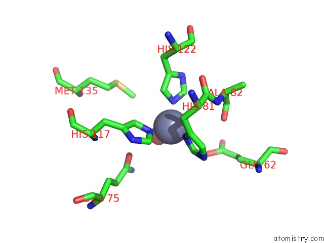
Mono view

Stereo pair view
Mono view
Stereo pair view
A full contact list of Zinc with other atoms in the Zn binding
site number 2 of Carbonic Anhydrase From Methanosarcina within 5.0Å range:
|
Zinc binding site 3 out of 3 in 1thj
Go back to
Zinc binding site 3 out
of 3 in the Carbonic Anhydrase From Methanosarcina
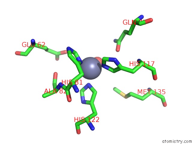
Mono view
Stereo pair view
Mono view
Stereo pair view
A full contact list of Zinc with other atoms in the Zn binding
site number 3 of Carbonic Anhydrase From Methanosarcina within 5.0Å range:
|
Reference:
C.Kisker,
H.Schindelin,
B.E.Alber,
J.G.Ferry,
D.C.Rees.
A Left-Hand Beta-Helix Revealed By the Crystal Structure of A Carbonic Anhydrase From the Archaeon Methanosarcina Thermophila. Embo J. V. 15 2323 1996.
ISSN: ISSN 0261-4189
PubMed: 8665839
Page generated: Wed Oct 16 19:08:28 2024
ISSN: ISSN 0261-4189
PubMed: 8665839
Last articles
Zn in 9J0NZn in 9J0O
Zn in 9J0P
Zn in 9FJX
Zn in 9EKB
Zn in 9C0F
Zn in 9CAH
Zn in 9CH0
Zn in 9CH3
Zn in 9CH1