Zinc »
PDB 1rrm-1sdy »
1s3g »
Zinc in PDB 1s3g: Crystal Structure of Adenylate Kinase From Bacillus Globisporus
Enzymatic activity of Crystal Structure of Adenylate Kinase From Bacillus Globisporus
All present enzymatic activity of Crystal Structure of Adenylate Kinase From Bacillus Globisporus:
2.7.4.3;
2.7.4.3;
Protein crystallography data
The structure of Crystal Structure of Adenylate Kinase From Bacillus Globisporus, PDB code: 1s3g
was solved by
E.Bae,
G.N.Phillips Jr.,
with X-Ray Crystallography technique. A brief refinement statistics is given in the table below:
| Resolution Low / High (Å) | 27.49 / 2.25 |
| Space group | P 31 2 1 |
| Cell size a, b, c (Å), α, β, γ (°) | 65.288, 65.288, 94.378, 90.00, 90.00, 120.00 |
| R / Rfree (%) | 22.3 / 28.8 |
Zinc Binding Sites:
The binding sites of Zinc atom in the Crystal Structure of Adenylate Kinase From Bacillus Globisporus
(pdb code 1s3g). This binding sites where shown within
5.0 Angstroms radius around Zinc atom.
In total only one binding site of Zinc was determined in the Crystal Structure of Adenylate Kinase From Bacillus Globisporus, PDB code: 1s3g:
In total only one binding site of Zinc was determined in the Crystal Structure of Adenylate Kinase From Bacillus Globisporus, PDB code: 1s3g:
Zinc binding site 1 out of 1 in 1s3g
Go back to
Zinc binding site 1 out
of 1 in the Crystal Structure of Adenylate Kinase From Bacillus Globisporus
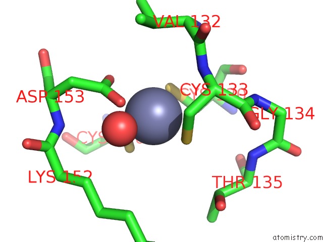
Mono view
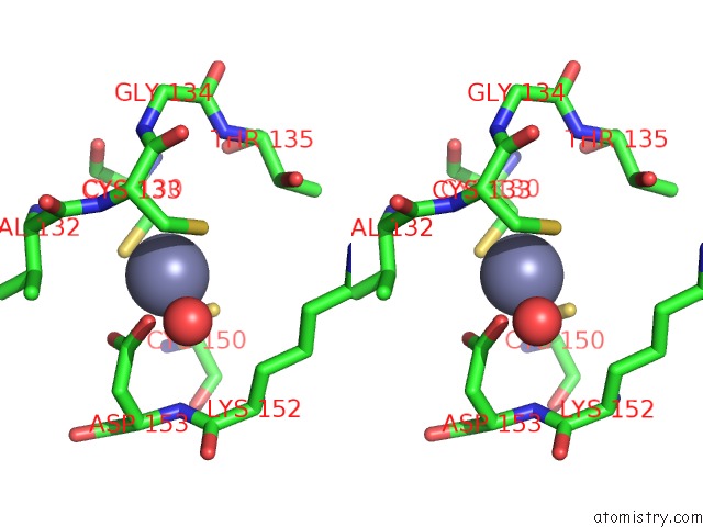
Stereo pair view
Mono view
Stereo pair view
A full contact list of Zinc with other atoms in the Zn binding
site number 1 of Crystal Structure of Adenylate Kinase From Bacillus Globisporus within 5.0Å range:
|
Reference:
E.Bae,
G.N.Phillips Jr..
Structures and Analysis of Highly Homologous Psychrophilic, Mesophilic, and Thermophilic Adenylate Kinases. J.Biol.Chem. V. 279 28202 2004.
ISSN: ISSN 0021-9258
PubMed: 15100224
DOI: 10.1074/JBC.M401865200
Page generated: Wed Oct 16 18:43:23 2024
ISSN: ISSN 0021-9258
PubMed: 15100224
DOI: 10.1074/JBC.M401865200
Last articles
Zn in 9J0NZn in 9J0O
Zn in 9J0P
Zn in 9FJX
Zn in 9EKB
Zn in 9C0F
Zn in 9CAH
Zn in 9CH0
Zn in 9CH3
Zn in 9CH1