Zinc »
PDB 1rrm-1sdy »
1rs7 »
Zinc in PDB 1rs7: Rat Neuronal Nos Heme Domain with D-Phenylalanine-D-Nitroarginine Amide Bound
Enzymatic activity of Rat Neuronal Nos Heme Domain with D-Phenylalanine-D-Nitroarginine Amide Bound
All present enzymatic activity of Rat Neuronal Nos Heme Domain with D-Phenylalanine-D-Nitroarginine Amide Bound:
1.14.13.39;
1.14.13.39;
Protein crystallography data
The structure of Rat Neuronal Nos Heme Domain with D-Phenylalanine-D-Nitroarginine Amide Bound, PDB code: 1rs7
was solved by
M.Flinspach,
H.Li,
J.Jamal,
W.Yang,
H.Huang,
R.B.Silverman,
T.L.Poulos,
with X-Ray Crystallography technique. A brief refinement statistics is given in the table below:
| Resolution Low / High (Å) | 39.06 / 1.95 |
| Space group | P 21 21 21 |
| Cell size a, b, c (Å), α, β, γ (°) | 52.085, 110.909, 165.032, 90.00, 90.00, 90.00 |
| R / Rfree (%) | 21.6 / 25.1 |
Other elements in 1rs7:
The structure of Rat Neuronal Nos Heme Domain with D-Phenylalanine-D-Nitroarginine Amide Bound also contains other interesting chemical elements:
| Iron | (Fe) | 2 atoms |
Zinc Binding Sites:
The binding sites of Zinc atom in the Rat Neuronal Nos Heme Domain with D-Phenylalanine-D-Nitroarginine Amide Bound
(pdb code 1rs7). This binding sites where shown within
5.0 Angstroms radius around Zinc atom.
In total only one binding site of Zinc was determined in the Rat Neuronal Nos Heme Domain with D-Phenylalanine-D-Nitroarginine Amide Bound, PDB code: 1rs7:
In total only one binding site of Zinc was determined in the Rat Neuronal Nos Heme Domain with D-Phenylalanine-D-Nitroarginine Amide Bound, PDB code: 1rs7:
Zinc binding site 1 out of 1 in 1rs7
Go back to
Zinc binding site 1 out
of 1 in the Rat Neuronal Nos Heme Domain with D-Phenylalanine-D-Nitroarginine Amide Bound
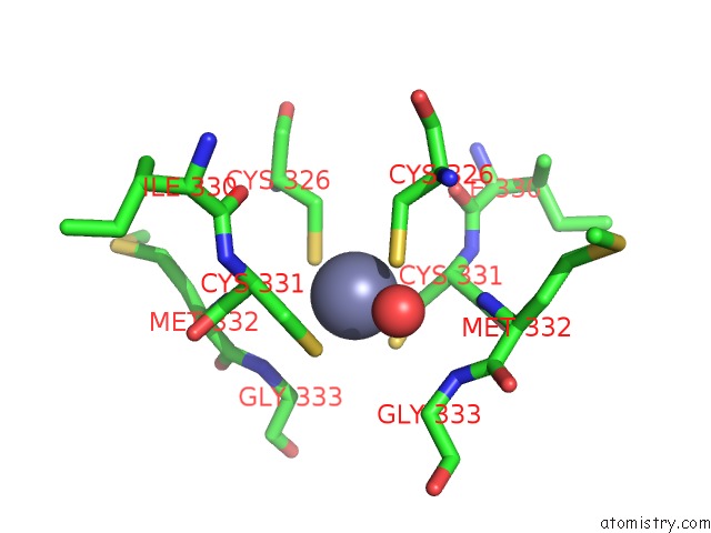
Mono view
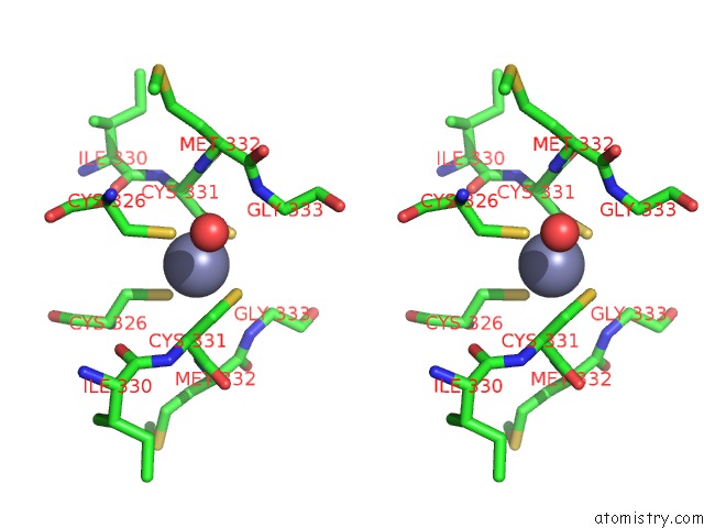
Stereo pair view
Mono view
Stereo pair view
A full contact list of Zinc with other atoms in the Zn binding
site number 1 of Rat Neuronal Nos Heme Domain with D-Phenylalanine-D-Nitroarginine Amide Bound within 5.0Å range:
|
Reference:
M.Flinspach,
H.Li,
J.Jamal,
W.Yang,
H.Huang,
R.B.Silverman,
T.L.Poulos.
Structures of the Neuronal and Endothelial Nitric Oxide Synthase Heme Domain with D-Nitroarginine-Containing Dipeptide Inhibitors Bound. Biochemistry V. 43 5181 2004.
ISSN: ISSN 0006-2960
PubMed: 15122883
DOI: 10.1021/BI0361867
Page generated: Wed Oct 16 18:39:56 2024
ISSN: ISSN 0006-2960
PubMed: 15122883
DOI: 10.1021/BI0361867
Last articles
Zn in 9J0NZn in 9J0O
Zn in 9J0P
Zn in 9FJX
Zn in 9EKB
Zn in 9C0F
Zn in 9CAH
Zn in 9CH0
Zn in 9CH3
Zn in 9CH1