Zinc »
PDB 1rag-1rqg »
1ro5 »
Zinc in PDB 1ro5: Crystal Structure of the Ahl Synthase Lasi
Protein crystallography data
The structure of Crystal Structure of the Ahl Synthase Lasi, PDB code: 1ro5
was solved by
T.A.Gould,
H.P.Schweizer,
M.E.Churchill,
with X-Ray Crystallography technique. A brief refinement statistics is given in the table below:
| Resolution Low / High (Å) | 35.58 / 2.30 |
| Space group | F 2 3 |
| Cell size a, b, c (Å), α, β, γ (°) | 154.900, 154.900, 154.900, 90.00, 90.00, 90.00 |
| R / Rfree (%) | 19.3 / 23.6 |
Zinc Binding Sites:
The binding sites of Zinc atom in the Crystal Structure of the Ahl Synthase Lasi
(pdb code 1ro5). This binding sites where shown within
5.0 Angstroms radius around Zinc atom.
In total only one binding site of Zinc was determined in the Crystal Structure of the Ahl Synthase Lasi, PDB code: 1ro5:
In total only one binding site of Zinc was determined in the Crystal Structure of the Ahl Synthase Lasi, PDB code: 1ro5:
Zinc binding site 1 out of 1 in 1ro5
Go back to
Zinc binding site 1 out
of 1 in the Crystal Structure of the Ahl Synthase Lasi
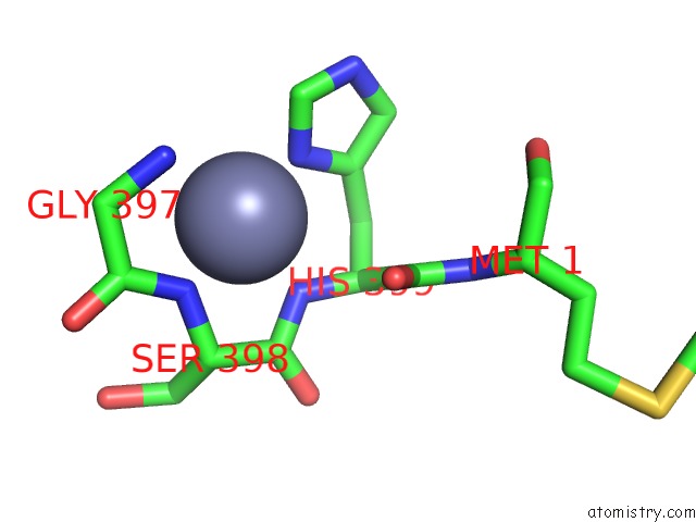
Mono view
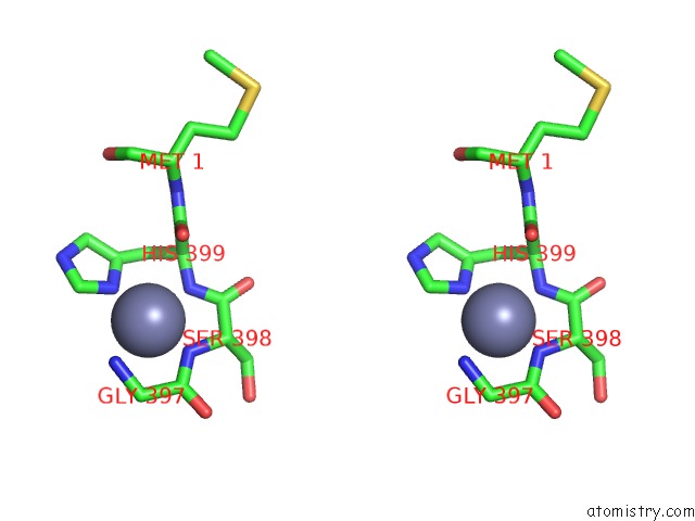
Stereo pair view
Mono view
Stereo pair view
A full contact list of Zinc with other atoms in the Zn binding
site number 1 of Crystal Structure of the Ahl Synthase Lasi within 5.0Å range:
|
Reference:
T.A.Gould,
H.P.Schweizer,
M.E.Churchill.
Structure of the Pseudomonas Aeruginosa Acyl-Homoserinelactone Synthase Lasi. Mol.Microbiol. V. 53 1135 2004.
ISSN: ISSN 0950-382X
PubMed: 15306017
DOI: 10.1111/J.1365-2958.2004.04211.X
Page generated: Wed Oct 16 18:37:51 2024
ISSN: ISSN 0950-382X
PubMed: 15306017
DOI: 10.1111/J.1365-2958.2004.04211.X
Last articles
Zn in 9J0NZn in 9J0O
Zn in 9J0P
Zn in 9FJX
Zn in 9EKB
Zn in 9C0F
Zn in 9CAH
Zn in 9CH0
Zn in 9CH3
Zn in 9CH1