Zinc »
PDB 1rag-1rqg »
1rfu »
Zinc in PDB 1rfu: Crystal Structure of Pyridoxal Kinase Complexed with Adp and Plp
Enzymatic activity of Crystal Structure of Pyridoxal Kinase Complexed with Adp and Plp
All present enzymatic activity of Crystal Structure of Pyridoxal Kinase Complexed with Adp and Plp:
2.7.1.35;
2.7.1.35;
Protein crystallography data
The structure of Crystal Structure of Pyridoxal Kinase Complexed with Adp and Plp, PDB code: 1rfu
was solved by
D.-C.Liang,
T.Jiang,
M.-H.Li,
with X-Ray Crystallography technique. A brief refinement statistics is given in the table below:
| Resolution Low / High (Å) | 20.00 / 2.80 |
| Space group | P 43 |
| Cell size a, b, c (Å), α, β, γ (°) | 109.088, 109.088, 284.272, 90.00, 90.00, 90.00 |
| R / Rfree (%) | 22.9 / 28.1 |
Zinc Binding Sites:
The binding sites of Zinc atom in the Crystal Structure of Pyridoxal Kinase Complexed with Adp and Plp
(pdb code 1rfu). This binding sites where shown within
5.0 Angstroms radius around Zinc atom.
In total 8 binding sites of Zinc where determined in the Crystal Structure of Pyridoxal Kinase Complexed with Adp and Plp, PDB code: 1rfu:
Jump to Zinc binding site number: 1; 2; 3; 4; 5; 6; 7; 8;
In total 8 binding sites of Zinc where determined in the Crystal Structure of Pyridoxal Kinase Complexed with Adp and Plp, PDB code: 1rfu:
Jump to Zinc binding site number: 1; 2; 3; 4; 5; 6; 7; 8;
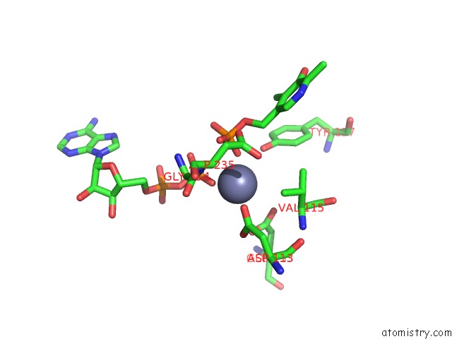
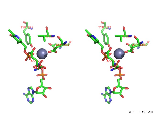
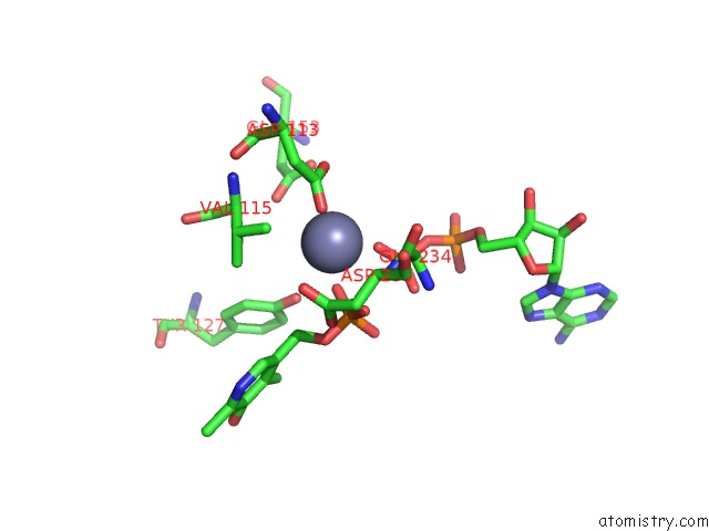
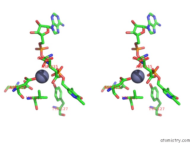
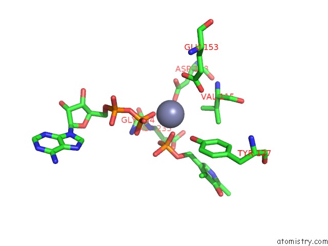
Zinc binding site 1 out of 8 in 1rfu
Go back to
Zinc binding site 1 out
of 8 in the Crystal Structure of Pyridoxal Kinase Complexed with Adp and Plp
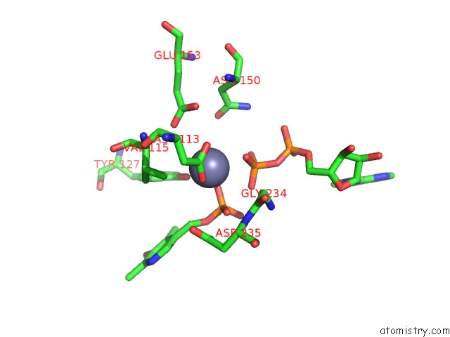
Mono view
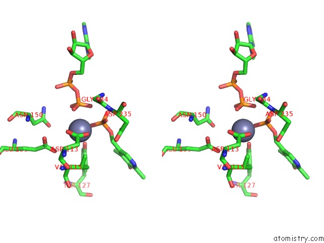
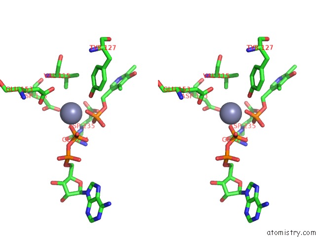
Stereo pair view
Mono view
Stereo pair view
A full contact list of Zinc with other atoms in the Zn binding
site number 1 of Crystal Structure of Pyridoxal Kinase Complexed with Adp and Plp within 5.0Å range:
|
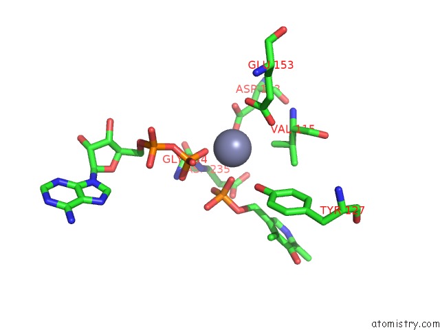
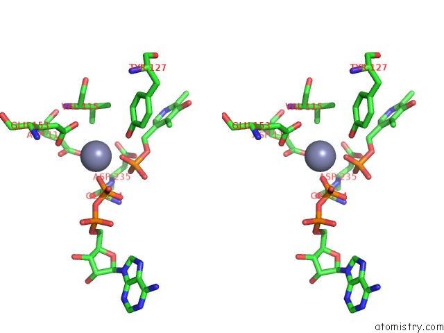
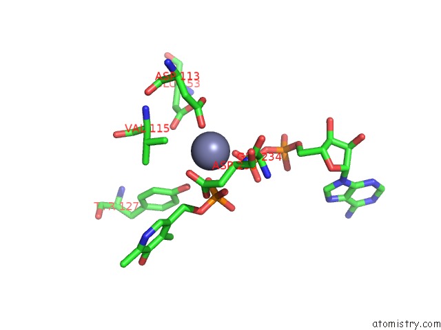
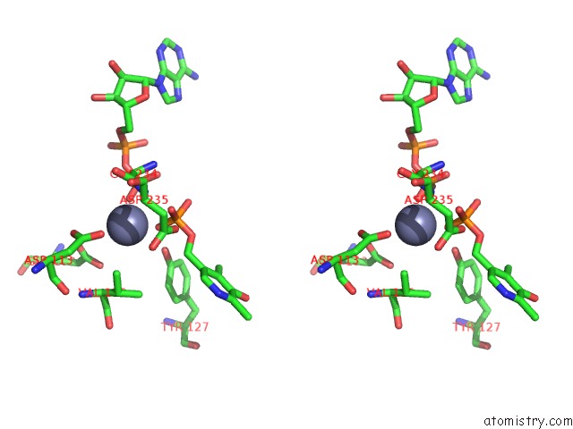
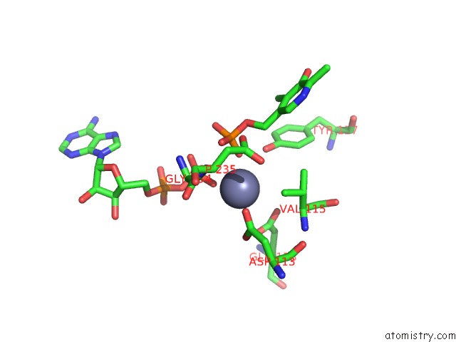
Zinc binding site 2 out of 8 in 1rfu
Go back to
Zinc binding site 2 out
of 8 in the Crystal Structure of Pyridoxal Kinase Complexed with Adp and Plp
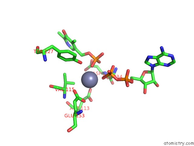
Mono view
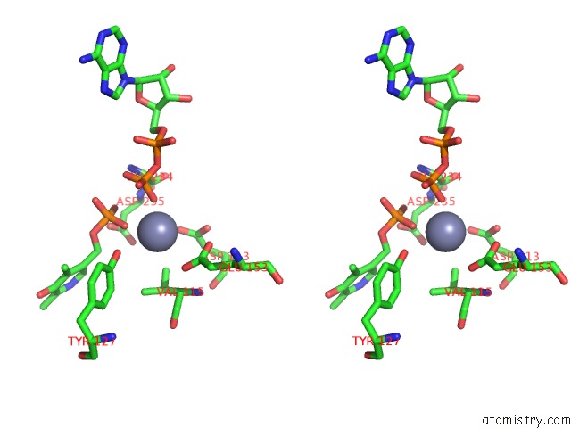
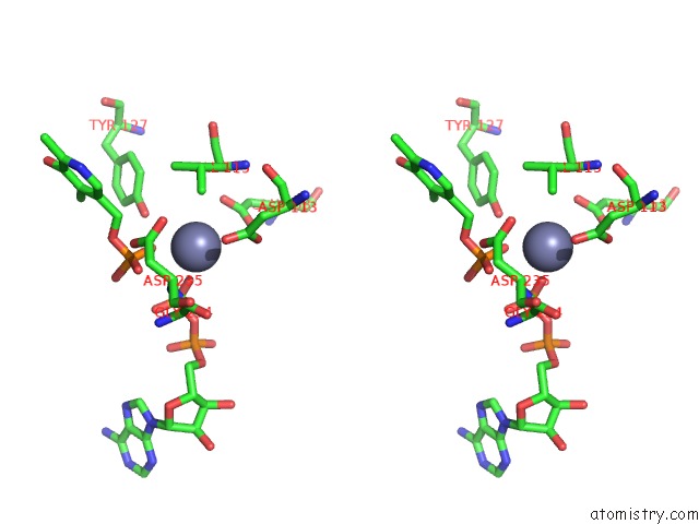
Stereo pair view
Mono view
Stereo pair view
A full contact list of Zinc with other atoms in the Zn binding
site number 2 of Crystal Structure of Pyridoxal Kinase Complexed with Adp and Plp within 5.0Å range:
|
Zinc binding site 3 out of 8 in 1rfu
Go back to
Zinc binding site 3 out
of 8 in the Crystal Structure of Pyridoxal Kinase Complexed with Adp and Plp
Mono view
Stereo pair view
Mono view
Stereo pair view
A full contact list of Zinc with other atoms in the Zn binding
site number 3 of Crystal Structure of Pyridoxal Kinase Complexed with Adp and Plp within 5.0Å range:
|
Zinc binding site 4 out of 8 in 1rfu
Go back to
Zinc binding site 4 out
of 8 in the Crystal Structure of Pyridoxal Kinase Complexed with Adp and Plp
Mono view
Stereo pair view
Mono view
Stereo pair view
A full contact list of Zinc with other atoms in the Zn binding
site number 4 of Crystal Structure of Pyridoxal Kinase Complexed with Adp and Plp within 5.0Å range:
|
Zinc binding site 5 out of 8 in 1rfu
Go back to
Zinc binding site 5 out
of 8 in the Crystal Structure of Pyridoxal Kinase Complexed with Adp and Plp
Mono view
Stereo pair view
Mono view
Stereo pair view
A full contact list of Zinc with other atoms in the Zn binding
site number 5 of Crystal Structure of Pyridoxal Kinase Complexed with Adp and Plp within 5.0Å range:
|
Zinc binding site 6 out of 8 in 1rfu
Go back to
Zinc binding site 6 out
of 8 in the Crystal Structure of Pyridoxal Kinase Complexed with Adp and Plp
Mono view
Stereo pair view
Mono view
Stereo pair view
A full contact list of Zinc with other atoms in the Zn binding
site number 6 of Crystal Structure of Pyridoxal Kinase Complexed with Adp and Plp within 5.0Å range:
|
Zinc binding site 7 out of 8 in 1rfu
Go back to
Zinc binding site 7 out
of 8 in the Crystal Structure of Pyridoxal Kinase Complexed with Adp and Plp
Mono view
Stereo pair view
Mono view
Stereo pair view
A full contact list of Zinc with other atoms in the Zn binding
site number 7 of Crystal Structure of Pyridoxal Kinase Complexed with Adp and Plp within 5.0Å range:
|
Zinc binding site 8 out of 8 in 1rfu
Go back to
Zinc binding site 8 out
of 8 in the Crystal Structure of Pyridoxal Kinase Complexed with Adp and Plp
Mono view
Stereo pair view
Mono view
Stereo pair view
A full contact list of Zinc with other atoms in the Zn binding
site number 8 of Crystal Structure of Pyridoxal Kinase Complexed with Adp and Plp within 5.0Å range:
|
Reference:
M.-H.Li,
F.Kwok,
W.-R.Chang,
S.-Q.Liu,
S.C.L.Lo,
J.-P.Zhang,
T.Jiang,
D.-C.Liang.
Conformational Changes in the Reaction of Pyridoxal Kinase J.Biol.Chem. V. 279 17459 2004.
ISSN: ISSN 0021-9258
PubMed: 14722069
DOI: 10.1074/JBC.M312380200
Page generated: Wed Oct 16 18:33:52 2024
ISSN: ISSN 0021-9258
PubMed: 14722069
DOI: 10.1074/JBC.M312380200
Last articles
Zn in 9J0NZn in 9J0O
Zn in 9J0P
Zn in 9FJX
Zn in 9EKB
Zn in 9C0F
Zn in 9CAH
Zn in 9CH0
Zn in 9CH3
Zn in 9CH1