Zinc »
PDB 1qmu-1r23 »
1r1i »
Zinc in PDB 1r1i: Structural Analysis of Neprilysin with Various Specific and Potent Inhibitors
Enzymatic activity of Structural Analysis of Neprilysin with Various Specific and Potent Inhibitors
All present enzymatic activity of Structural Analysis of Neprilysin with Various Specific and Potent Inhibitors:
3.4.24.11;
3.4.24.11;
Protein crystallography data
The structure of Structural Analysis of Neprilysin with Various Specific and Potent Inhibitors, PDB code: 1r1i
was solved by
C.Oefner,
B.P.Roques,
M.C.Fournie-Zaluski,
G.E.Dale,
with X-Ray Crystallography technique. A brief refinement statistics is given in the table below:
| Resolution Low / High (Å) | 20.00 / 2.60 |
| Space group | P 32 2 1 |
| Cell size a, b, c (Å), α, β, γ (°) | 108.845, 108.845, 113.071, 90.00, 90.00, 120.00 |
| R / Rfree (%) | 27.6 / 35.8 |
Zinc Binding Sites:
The binding sites of Zinc atom in the Structural Analysis of Neprilysin with Various Specific and Potent Inhibitors
(pdb code 1r1i). This binding sites where shown within
5.0 Angstroms radius around Zinc atom.
In total only one binding site of Zinc was determined in the Structural Analysis of Neprilysin with Various Specific and Potent Inhibitors, PDB code: 1r1i:
In total only one binding site of Zinc was determined in the Structural Analysis of Neprilysin with Various Specific and Potent Inhibitors, PDB code: 1r1i:
Zinc binding site 1 out of 1 in 1r1i
Go back to
Zinc binding site 1 out
of 1 in the Structural Analysis of Neprilysin with Various Specific and Potent Inhibitors
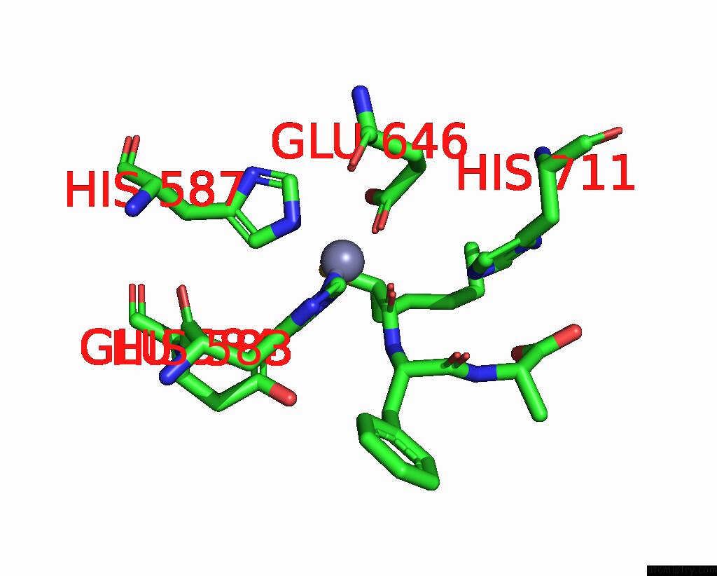
Mono view
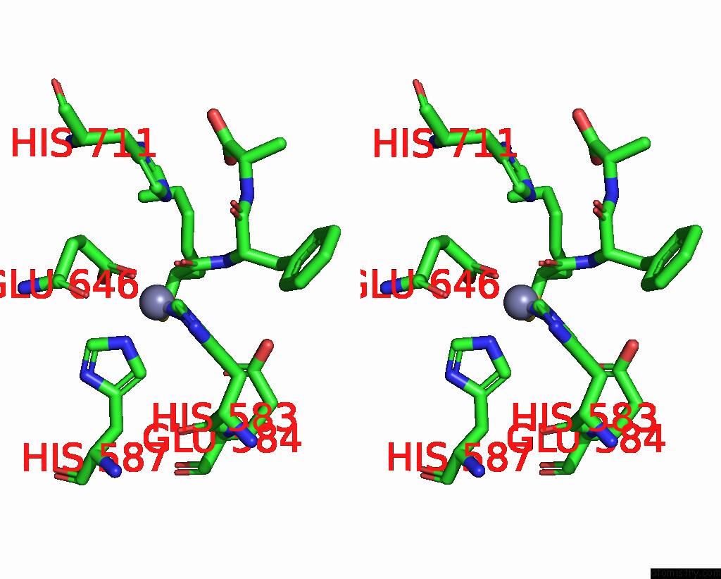
Stereo pair view
Mono view
Stereo pair view
A full contact list of Zinc with other atoms in the Zn binding
site number 1 of Structural Analysis of Neprilysin with Various Specific and Potent Inhibitors within 5.0Å range:
|
Reference:
C.Oefner,
B.P.Roques,
M.C.Fournie-Zaluski,
G.E.Dale.
Structural Analysis of Neprilysin with Various Specific and Potent Inhibitors. Acta Crystallogr.,Sect.D V. 60 392 2004.
ISSN: ISSN 0907-4449
PubMed: 14747736
DOI: 10.1107/S0907444903027410
Page generated: Wed Oct 16 18:19:38 2024
ISSN: ISSN 0907-4449
PubMed: 14747736
DOI: 10.1107/S0907444903027410
Last articles
Zn in 9J0NZn in 9J0O
Zn in 9J0P
Zn in 9FJX
Zn in 9EKB
Zn in 9C0F
Zn in 9CAH
Zn in 9CH0
Zn in 9CH3
Zn in 9CH1