Zinc »
PDB 1qmu-1r23 »
1qtw »
Zinc in PDB 1qtw: High-Resolution Crystal Structure of the Escherichia Coli Dna Repair Enzyme Endonuclease IV
Enzymatic activity of High-Resolution Crystal Structure of the Escherichia Coli Dna Repair Enzyme Endonuclease IV
All present enzymatic activity of High-Resolution Crystal Structure of the Escherichia Coli Dna Repair Enzyme Endonuclease IV:
3.1.21.2;
3.1.21.2;
Protein crystallography data
The structure of High-Resolution Crystal Structure of the Escherichia Coli Dna Repair Enzyme Endonuclease IV, PDB code: 1qtw
was solved by
D.J.Hosfield,
Y.Guan,
B.J.Haas,
R.P.Cunningham,
J.A.Tainer,
with X-Ray Crystallography technique. A brief refinement statistics is given in the table below:
| Resolution Low / High (Å) | 20.00 / 1.02 |
| Space group | P 1 21 1 |
| Cell size a, b, c (Å), α, β, γ (°) | 49.620, 59.560, 50.980, 90.00, 110.94, 90.00 |
| R / Rfree (%) | 12.4 / 14.8 |
Zinc Binding Sites:
The binding sites of Zinc atom in the High-Resolution Crystal Structure of the Escherichia Coli Dna Repair Enzyme Endonuclease IV
(pdb code 1qtw). This binding sites where shown within
5.0 Angstroms radius around Zinc atom.
In total 3 binding sites of Zinc where determined in the High-Resolution Crystal Structure of the Escherichia Coli Dna Repair Enzyme Endonuclease IV, PDB code: 1qtw:
Jump to Zinc binding site number: 1; 2; 3;
In total 3 binding sites of Zinc where determined in the High-Resolution Crystal Structure of the Escherichia Coli Dna Repair Enzyme Endonuclease IV, PDB code: 1qtw:
Jump to Zinc binding site number: 1; 2; 3;
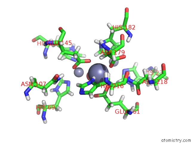
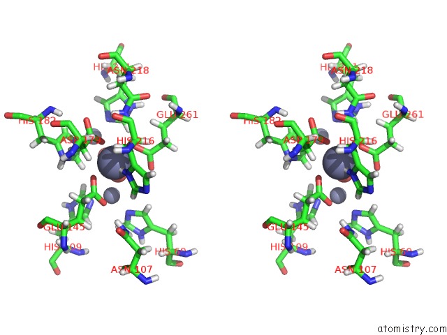
Zinc binding site 1 out of 3 in 1qtw
Go back to
Zinc binding site 1 out
of 3 in the High-Resolution Crystal Structure of the Escherichia Coli Dna Repair Enzyme Endonuclease IV
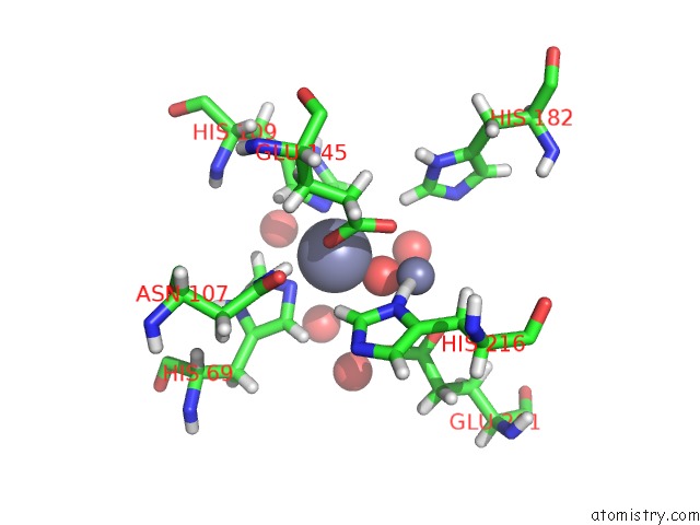
Mono view
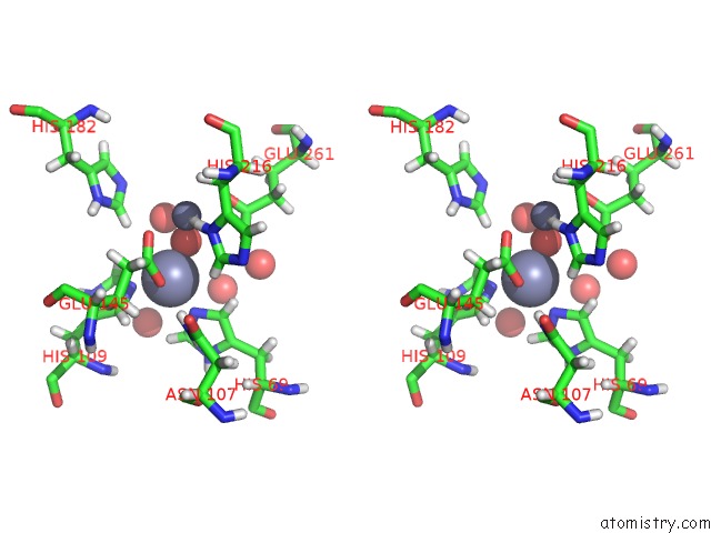
Stereo pair view
Mono view
Stereo pair view
A full contact list of Zinc with other atoms in the Zn binding
site number 1 of High-Resolution Crystal Structure of the Escherichia Coli Dna Repair Enzyme Endonuclease IV within 5.0Å range:
|
Zinc binding site 2 out of 3 in 1qtw
Go back to
Zinc binding site 2 out
of 3 in the High-Resolution Crystal Structure of the Escherichia Coli Dna Repair Enzyme Endonuclease IV
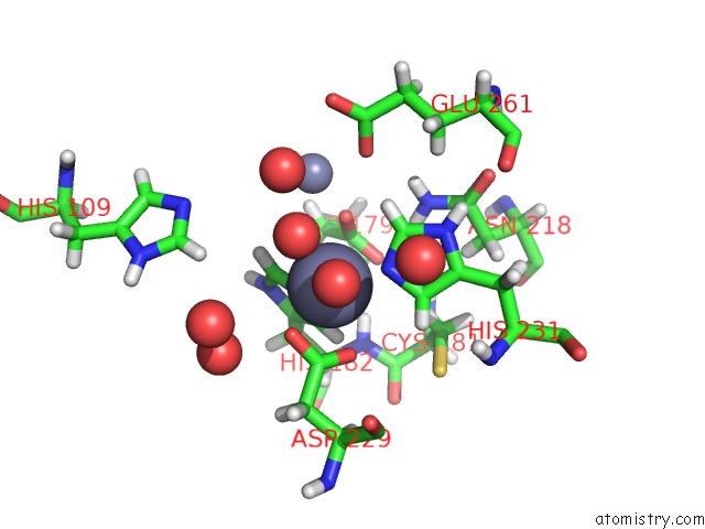
Mono view
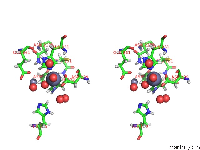
Stereo pair view
Mono view
Stereo pair view
A full contact list of Zinc with other atoms in the Zn binding
site number 2 of High-Resolution Crystal Structure of the Escherichia Coli Dna Repair Enzyme Endonuclease IV within 5.0Å range:
|
Zinc binding site 3 out of 3 in 1qtw
Go back to
Zinc binding site 3 out
of 3 in the High-Resolution Crystal Structure of the Escherichia Coli Dna Repair Enzyme Endonuclease IV
Mono view
Stereo pair view
Mono view
Stereo pair view
A full contact list of Zinc with other atoms in the Zn binding
site number 3 of High-Resolution Crystal Structure of the Escherichia Coli Dna Repair Enzyme Endonuclease IV within 5.0Å range:
|
Reference:
D.J.Hosfield,
Y.Guan,
B.J.Haas,
R.P.Cunningham,
J.A.Tainer.
Structure of the Dna Repair Enzyme Endonuclease IV and Its Dna Complex: Double-Nucleotide Flipping at Abasic Sites and Three-Metal-Ion Catalysis. Cell(Cambridge,Mass.) V. 98 397 1999.
ISSN: ISSN 0092-8674
PubMed: 10458614
DOI: 10.1016/S0092-8674(00)81968-6
Page generated: Wed Oct 16 18:15:13 2024
ISSN: ISSN 0092-8674
PubMed: 10458614
DOI: 10.1016/S0092-8674(00)81968-6
Last articles
Zn in 9J0NZn in 9J0O
Zn in 9J0P
Zn in 9FJX
Zn in 9EKB
Zn in 9C0F
Zn in 9CAH
Zn in 9CH0
Zn in 9CH3
Zn in 9CH1