Zinc »
PDB 1q66-1qmd »
1qiy »
Zinc in PDB 1qiy: Human Insulin Hexamers with Chain B His Mutated to Tyr Complexed with Phenol
Protein crystallography data
The structure of Human Insulin Hexamers with Chain B His Mutated to Tyr Complexed with Phenol, PDB code: 1qiy
was solved by
L.Tang,
J.L.Whittingham,
C.S.Verma,
L.S.D.Caves,
G.G.Dodson,
with X-Ray Crystallography technique. A brief refinement statistics is given in the table below:
| Resolution Low / High (Å) | 36.80 / 2.30 |
| Space group | P 1 21 1 |
| Cell size a, b, c (Å), α, β, γ (°) | 61.100, 62.080, 48.350, 90.00, 109.87, 90.00 |
| R / Rfree (%) | 18.6 / n/a |
Other elements in 1qiy:
The structure of Human Insulin Hexamers with Chain B His Mutated to Tyr Complexed with Phenol also contains other interesting chemical elements:
| Chlorine | (Cl) | 2 atoms |
Zinc Binding Sites:
The binding sites of Zinc atom in the Human Insulin Hexamers with Chain B His Mutated to Tyr Complexed with Phenol
(pdb code 1qiy). This binding sites where shown within
5.0 Angstroms radius around Zinc atom.
In total 2 binding sites of Zinc where determined in the Human Insulin Hexamers with Chain B His Mutated to Tyr Complexed with Phenol, PDB code: 1qiy:
Jump to Zinc binding site number: 1; 2;
In total 2 binding sites of Zinc where determined in the Human Insulin Hexamers with Chain B His Mutated to Tyr Complexed with Phenol, PDB code: 1qiy:
Jump to Zinc binding site number: 1; 2;
Zinc binding site 1 out of 2 in 1qiy
Go back to
Zinc binding site 1 out
of 2 in the Human Insulin Hexamers with Chain B His Mutated to Tyr Complexed with Phenol
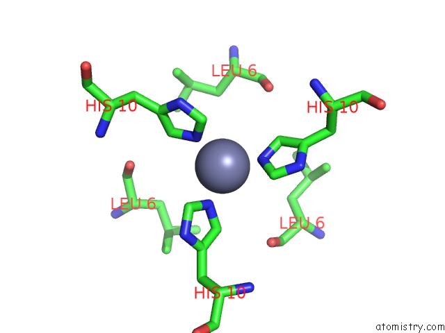
Mono view
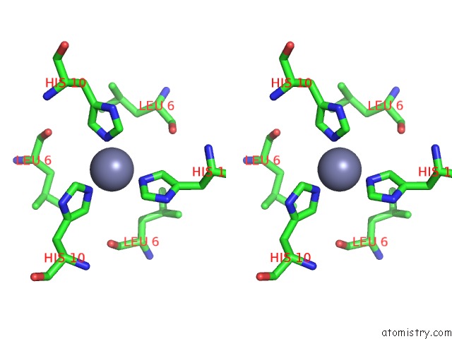
Stereo pair view
Mono view
Stereo pair view
A full contact list of Zinc with other atoms in the Zn binding
site number 1 of Human Insulin Hexamers with Chain B His Mutated to Tyr Complexed with Phenol within 5.0Å range:
|
Zinc binding site 2 out of 2 in 1qiy
Go back to
Zinc binding site 2 out
of 2 in the Human Insulin Hexamers with Chain B His Mutated to Tyr Complexed with Phenol
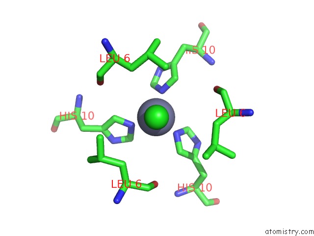
Mono view
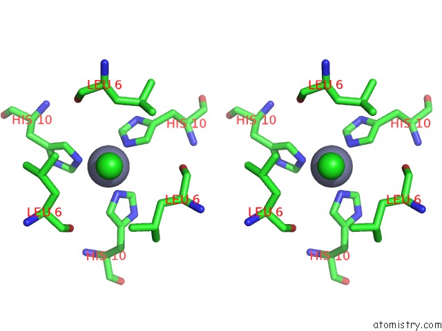
Stereo pair view
Mono view
Stereo pair view
A full contact list of Zinc with other atoms in the Zn binding
site number 2 of Human Insulin Hexamers with Chain B His Mutated to Tyr Complexed with Phenol within 5.0Å range:
|
Reference:
L.Tang,
J.L.Whittingham,
C.S.Verma,
L.S.D.Caves,
G.G.Dodson.
Structural Consequences of the B5 Histidine --> Tyrosine Mutation in Human Insulin Characterized By X-Ray Crystallography and Conformational Analysis. Biochemistry V. 38 12041 1999.
ISSN: ISSN 0006-2960
PubMed: 10508408
DOI: 10.1021/BI990700K
Page generated: Wed Oct 16 18:11:24 2024
ISSN: ISSN 0006-2960
PubMed: 10508408
DOI: 10.1021/BI990700K
Last articles
Zn in 9J0NZn in 9J0O
Zn in 9J0P
Zn in 9FJX
Zn in 9EKB
Zn in 9C0F
Zn in 9CAH
Zn in 9CH0
Zn in 9CH3
Zn in 9CH1