Zinc »
PDB 1q66-1qmd »
1qbq »
Zinc in PDB 1qbq: Structure of Rat Farnesyl Protein Transferase Complexed with A Cvim Peptide and Alpha-Hydroxyfarnesylphosphonic Acid.
Protein crystallography data
The structure of Structure of Rat Farnesyl Protein Transferase Complexed with A Cvim Peptide and Alpha-Hydroxyfarnesylphosphonic Acid., PDB code: 1qbq
was solved by
C.L.Strickland,
W.T.Windsor,
R.Syto,
L.Wang,
R.Bond,
Z.Wu,
J.Schwartz,
H.V.Le,
L.S.Beese,
P.C.Weber,
with X-Ray Crystallography technique. A brief refinement statistics is given in the table below:
| Resolution Low / High (Å) | 15.00 / 2.40 |
| Space group | P 61 |
| Cell size a, b, c (Å), α, β, γ (°) | 174.132, 174.132, 69.705, 90.00, 90.00, 120.00 |
| R / Rfree (%) | 21.8 / 29.2 |
Zinc Binding Sites:
The binding sites of Zinc atom in the Structure of Rat Farnesyl Protein Transferase Complexed with A Cvim Peptide and Alpha-Hydroxyfarnesylphosphonic Acid.
(pdb code 1qbq). This binding sites where shown within
5.0 Angstroms radius around Zinc atom.
In total only one binding site of Zinc was determined in the Structure of Rat Farnesyl Protein Transferase Complexed with A Cvim Peptide and Alpha-Hydroxyfarnesylphosphonic Acid., PDB code: 1qbq:
In total only one binding site of Zinc was determined in the Structure of Rat Farnesyl Protein Transferase Complexed with A Cvim Peptide and Alpha-Hydroxyfarnesylphosphonic Acid., PDB code: 1qbq:
Zinc binding site 1 out of 1 in 1qbq
Go back to
Zinc binding site 1 out
of 1 in the Structure of Rat Farnesyl Protein Transferase Complexed with A Cvim Peptide and Alpha-Hydroxyfarnesylphosphonic Acid.
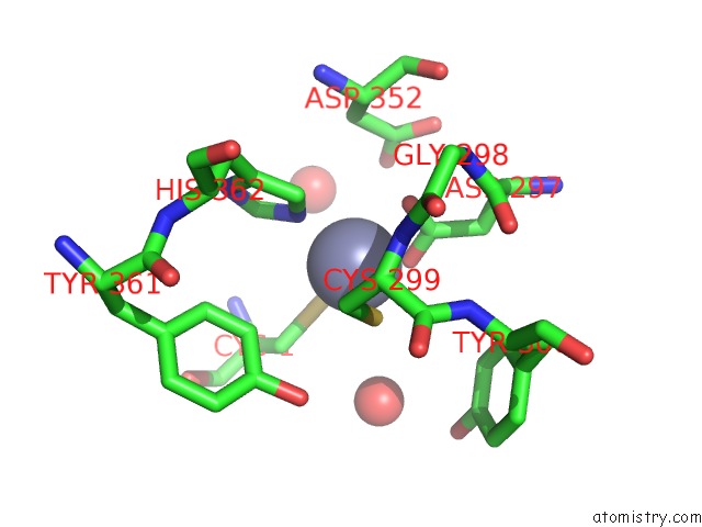
Mono view
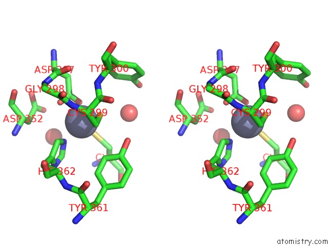
Stereo pair view
Mono view
Stereo pair view
A full contact list of Zinc with other atoms in the Zn binding
site number 1 of Structure of Rat Farnesyl Protein Transferase Complexed with A Cvim Peptide and Alpha-Hydroxyfarnesylphosphonic Acid. within 5.0Å range:
|
Reference:
C.L.Strickland,
W.T.Windsor,
R.Syto,
L.Wang,
R.Bond,
Z.Wu,
J.Schwartz,
H.V.Le,
L.S.Beese,
P.C.Weber.
Crystal Structure of Farnesyl Protein Transferase Complexed with A Caax Peptide and Farnesyl Diphosphate Analogue Biochemistry V. 37 16601 1998.
ISSN: ISSN 0006-2960
PubMed: 9843427
DOI: 10.1021/BI981197Z
Page generated: Wed Oct 16 18:07:35 2024
ISSN: ISSN 0006-2960
PubMed: 9843427
DOI: 10.1021/BI981197Z
Last articles
Zn in 9J0NZn in 9J0O
Zn in 9J0P
Zn in 9FJX
Zn in 9EKB
Zn in 9C0F
Zn in 9CAH
Zn in 9CH0
Zn in 9CH3
Zn in 9CH1