Zinc »
PDB 1q66-1qmd »
1q74 »
Zinc in PDB 1q74: The Crystal Structure of 1D-Myo-Inosityl 2-Acetamido-2- Deoxy-Alpha-D-Glucopyranoside Deacetylase (Mshb)
Protein crystallography data
The structure of The Crystal Structure of 1D-Myo-Inosityl 2-Acetamido-2- Deoxy-Alpha-D-Glucopyranoside Deacetylase (Mshb), PDB code: 1q74
was solved by
J.T.Maynes,
C.Garen,
M.M.Cherney,
G.Newton,
D.Arad,
Y.Av-Gay,
R.C.Fahey,
M.N.James,
Tb Structural Genomics Consortium(Tbsgc),
with X-Ray Crystallography technique. A brief refinement statistics is given in the table below:
| Resolution Low / High (Å) | 19.84 / 1.70 |
| Space group | P 1 |
| Cell size a, b, c (Å), α, β, γ (°) | 56.794, 73.964, 85.587, 102.09, 108.16, 97.18 |
| R / Rfree (%) | 19.4 / 22.8 |
Zinc Binding Sites:
The binding sites of Zinc atom in the The Crystal Structure of 1D-Myo-Inosityl 2-Acetamido-2- Deoxy-Alpha-D-Glucopyranoside Deacetylase (Mshb)
(pdb code 1q74). This binding sites where shown within
5.0 Angstroms radius around Zinc atom.
In total 4 binding sites of Zinc where determined in the The Crystal Structure of 1D-Myo-Inosityl 2-Acetamido-2- Deoxy-Alpha-D-Glucopyranoside Deacetylase (Mshb), PDB code: 1q74:
Jump to Zinc binding site number: 1; 2; 3; 4;
In total 4 binding sites of Zinc where determined in the The Crystal Structure of 1D-Myo-Inosityl 2-Acetamido-2- Deoxy-Alpha-D-Glucopyranoside Deacetylase (Mshb), PDB code: 1q74:
Jump to Zinc binding site number: 1; 2; 3; 4;
Zinc binding site 1 out of 4 in 1q74
Go back to
Zinc binding site 1 out
of 4 in the The Crystal Structure of 1D-Myo-Inosityl 2-Acetamido-2- Deoxy-Alpha-D-Glucopyranoside Deacetylase (Mshb)

Mono view
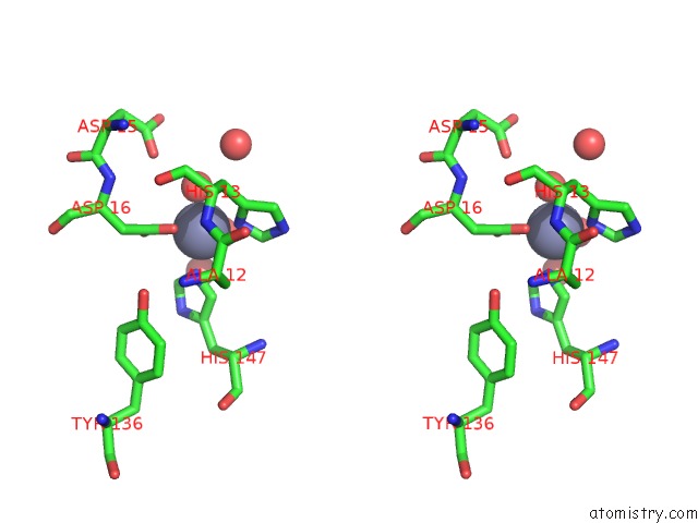
Stereo pair view
Mono view
Stereo pair view
A full contact list of Zinc with other atoms in the Zn binding
site number 1 of The Crystal Structure of 1D-Myo-Inosityl 2-Acetamido-2- Deoxy-Alpha-D-Glucopyranoside Deacetylase (Mshb) within 5.0Å range:
|
Zinc binding site 2 out of 4 in 1q74
Go back to
Zinc binding site 2 out
of 4 in the The Crystal Structure of 1D-Myo-Inosityl 2-Acetamido-2- Deoxy-Alpha-D-Glucopyranoside Deacetylase (Mshb)
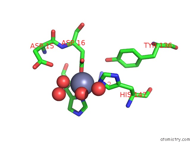
Mono view

Stereo pair view
Mono view
Stereo pair view
A full contact list of Zinc with other atoms in the Zn binding
site number 2 of The Crystal Structure of 1D-Myo-Inosityl 2-Acetamido-2- Deoxy-Alpha-D-Glucopyranoside Deacetylase (Mshb) within 5.0Å range:
|
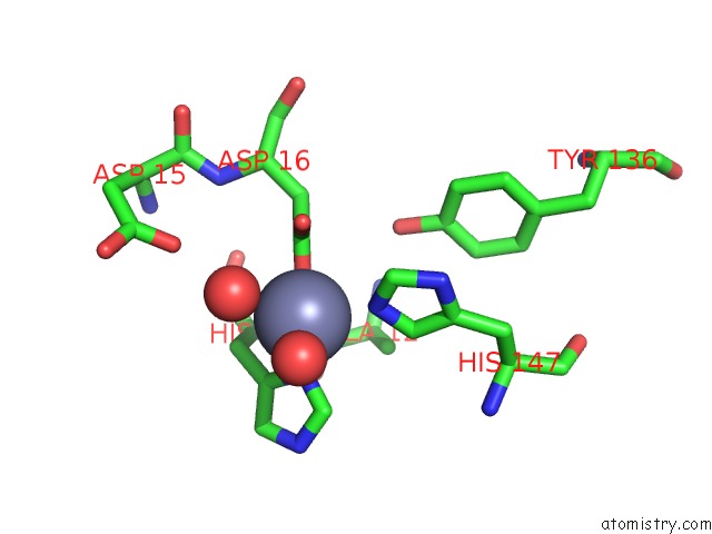
Zinc binding site 3 out of 4 in 1q74
Go back to
Zinc binding site 3 out
of 4 in the The Crystal Structure of 1D-Myo-Inosityl 2-Acetamido-2- Deoxy-Alpha-D-Glucopyranoside Deacetylase (Mshb)

Mono view

Stereo pair view
Mono view
Stereo pair view
A full contact list of Zinc with other atoms in the Zn binding
site number 3 of The Crystal Structure of 1D-Myo-Inosityl 2-Acetamido-2- Deoxy-Alpha-D-Glucopyranoside Deacetylase (Mshb) within 5.0Å range:
|
Zinc binding site 4 out of 4 in 1q74
Go back to
Zinc binding site 4 out
of 4 in the The Crystal Structure of 1D-Myo-Inosityl 2-Acetamido-2- Deoxy-Alpha-D-Glucopyranoside Deacetylase (Mshb)
Mono view

Stereo pair view
Mono view
Stereo pair view
A full contact list of Zinc with other atoms in the Zn binding
site number 4 of The Crystal Structure of 1D-Myo-Inosityl 2-Acetamido-2- Deoxy-Alpha-D-Glucopyranoside Deacetylase (Mshb) within 5.0Å range:
|
Reference:
J.T.Maynes,
C.Garen,
M.M.Cherney,
G.Newton,
D.Arad,
Y.Av-Gay,
R.C.Fahey,
M.N.James.
The Crystal Structure of 1-D-Myo-Inosityl 2-Acetamido-2-Deoxy-Alpha-D-Glucopyranoside Deacetylase (Mshb) From Mycobacterium Tuberculosis Reveals A Zinc Hydrolase with A Lactate Dehydrogenase Fold. J.Biol.Chem. V. 278 47166 2003.
ISSN: ISSN 0021-9258
PubMed: 12958317
DOI: 10.1074/JBC.M308914200
Page generated: Wed Oct 16 18:05:41 2024
ISSN: ISSN 0021-9258
PubMed: 12958317
DOI: 10.1074/JBC.M308914200
Last articles
Zn in 9J0NZn in 9J0O
Zn in 9J0P
Zn in 9FJX
Zn in 9EKB
Zn in 9C0F
Zn in 9CAH
Zn in 9CH0
Zn in 9CH3
Zn in 9CH1