Zinc »
PDB 1q66-1qmd »
1q66 »
Zinc in PDB 1q66: Crystal Structure of Tgt in Complex with 2-Amino-6-Aminomethyl-8- Phenylsulfanylmethyl-3H-Quinazolin-4-One Crystallized at pH 5.5
Enzymatic activity of Crystal Structure of Tgt in Complex with 2-Amino-6-Aminomethyl-8- Phenylsulfanylmethyl-3H-Quinazolin-4-One Crystallized at pH 5.5
All present enzymatic activity of Crystal Structure of Tgt in Complex with 2-Amino-6-Aminomethyl-8- Phenylsulfanylmethyl-3H-Quinazolin-4-One Crystallized at pH 5.5:
2.4.2.29;
2.4.2.29;
Protein crystallography data
The structure of Crystal Structure of Tgt in Complex with 2-Amino-6-Aminomethyl-8- Phenylsulfanylmethyl-3H-Quinazolin-4-One Crystallized at pH 5.5, PDB code: 1q66
was solved by
R.Brenk,
E.Meyer,
K.Reuter,
M.T.Stubbs,
G.A.Garcia,
G.Klebe,
with X-Ray Crystallography technique. A brief refinement statistics is given in the table below:
| Resolution Low / High (Å) | 40.00 / 1.75 |
| Space group | C 1 2 1 |
| Cell size a, b, c (Å), α, β, γ (°) | 91.400, 65.090, 69.850, 90.00, 95.85, 90.00 |
| R / Rfree (%) | 18.9 / 21 |
Zinc Binding Sites:
The binding sites of Zinc atom in the Crystal Structure of Tgt in Complex with 2-Amino-6-Aminomethyl-8- Phenylsulfanylmethyl-3H-Quinazolin-4-One Crystallized at pH 5.5
(pdb code 1q66). This binding sites where shown within
5.0 Angstroms radius around Zinc atom.
In total only one binding site of Zinc was determined in the Crystal Structure of Tgt in Complex with 2-Amino-6-Aminomethyl-8- Phenylsulfanylmethyl-3H-Quinazolin-4-One Crystallized at pH 5.5, PDB code: 1q66:
In total only one binding site of Zinc was determined in the Crystal Structure of Tgt in Complex with 2-Amino-6-Aminomethyl-8- Phenylsulfanylmethyl-3H-Quinazolin-4-One Crystallized at pH 5.5, PDB code: 1q66:
Zinc binding site 1 out of 1 in 1q66
Go back to
Zinc binding site 1 out
of 1 in the Crystal Structure of Tgt in Complex with 2-Amino-6-Aminomethyl-8- Phenylsulfanylmethyl-3H-Quinazolin-4-One Crystallized at pH 5.5
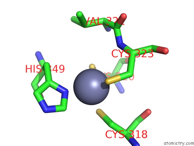
Mono view

Stereo pair view
Mono view
Stereo pair view
A full contact list of Zinc with other atoms in the Zn binding
site number 1 of Crystal Structure of Tgt in Complex with 2-Amino-6-Aminomethyl-8- Phenylsulfanylmethyl-3H-Quinazolin-4-One Crystallized at pH 5.5 within 5.0Å range:
|
Reference:
R.Brenk,
E.Meyer,
K.Reuter,
M.T.Stubbs,
G.A.Garcia,
F.Diederich,
G.Klebe.
Crystallographic Study of Inhibitors of Trna-Guanine Transglycosylase Suggests A New Structure-Based Pharmacophore For Virtual Screening. J.Mol.Biol. V. 338 55 2004.
ISSN: ISSN 0022-2836
PubMed: 15050823
DOI: 10.1016/J.JMB.2004.02.019
Page generated: Wed Oct 16 18:05:20 2024
ISSN: ISSN 0022-2836
PubMed: 15050823
DOI: 10.1016/J.JMB.2004.02.019
Last articles
Zn in 9J0NZn in 9J0O
Zn in 9J0P
Zn in 9FJX
Zn in 9EKB
Zn in 9C0F
Zn in 9CAH
Zn in 9CH0
Zn in 9CH3
Zn in 9CH1