Zinc »
PDB 1pvw-1q65 »
1q1y »
Zinc in PDB 1q1y: Crystal Structures of Peptide Deformylase From Staphylococcus Aureus Complexed with Actinonin
Enzymatic activity of Crystal Structures of Peptide Deformylase From Staphylococcus Aureus Complexed with Actinonin
All present enzymatic activity of Crystal Structures of Peptide Deformylase From Staphylococcus Aureus Complexed with Actinonin:
3.5.1.88;
3.5.1.88;
Protein crystallography data
The structure of Crystal Structures of Peptide Deformylase From Staphylococcus Aureus Complexed with Actinonin, PDB code: 1q1y
was solved by
H.J.Yoon,
S.K.Lee,
H.L.Kim,
H.W.Kim,
H.W.Kim,
J.Y.Lee,
B.Mikami,
S.W.Suh,
with X-Ray Crystallography technique. A brief refinement statistics is given in the table below:
| Resolution Low / High (Å) | 19.99 / 1.90 |
| Space group | C 2 2 21 |
| Cell size a, b, c (Å), α, β, γ (°) | 94.417, 120.844, 48.057, 90.00, 90.00, 90.00 |
| R / Rfree (%) | 20.5 / 23.5 |
Zinc Binding Sites:
The binding sites of Zinc atom in the Crystal Structures of Peptide Deformylase From Staphylococcus Aureus Complexed with Actinonin
(pdb code 1q1y). This binding sites where shown within
5.0 Angstroms radius around Zinc atom.
In total only one binding site of Zinc was determined in the Crystal Structures of Peptide Deformylase From Staphylococcus Aureus Complexed with Actinonin, PDB code: 1q1y:
In total only one binding site of Zinc was determined in the Crystal Structures of Peptide Deformylase From Staphylococcus Aureus Complexed with Actinonin, PDB code: 1q1y:
Zinc binding site 1 out of 1 in 1q1y
Go back to
Zinc binding site 1 out
of 1 in the Crystal Structures of Peptide Deformylase From Staphylococcus Aureus Complexed with Actinonin
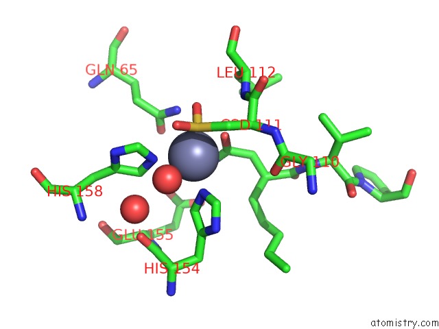
Mono view
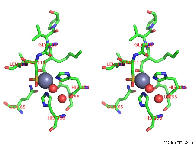
Stereo pair view
Mono view
Stereo pair view
A full contact list of Zinc with other atoms in the Zn binding
site number 1 of Crystal Structures of Peptide Deformylase From Staphylococcus Aureus Complexed with Actinonin within 5.0Å range:
|
Reference:
H.J.Yoon,
H.L.Kim,
S.K.Lee,
H.W.Kim,
H.W.Kim,
J.Y.Lee,
B.Mikami,
S.W.Suh.
Crystal Structure of Peptide Deformylase From Staphylococcus Aureus in Complex with Actinonin, A Naturally Occurring Antibacterial Agent Proteins V. 57 639 2004.
ISSN: ISSN 0887-3585
PubMed: 15382235
DOI: 10.1002/PROT.20231
Page generated: Wed Oct 16 18:00:34 2024
ISSN: ISSN 0887-3585
PubMed: 15382235
DOI: 10.1002/PROT.20231
Last articles
Zn in 9J0NZn in 9J0O
Zn in 9J0P
Zn in 9FJX
Zn in 9EKB
Zn in 9C0F
Zn in 9CAH
Zn in 9CH0
Zn in 9CH3
Zn in 9CH1