Zinc »
PDB 1peg-1pv9 »
1ptm »
Zinc in PDB 1ptm: Crystal Structure of E.Coli Pdxa
Enzymatic activity of Crystal Structure of E.Coli Pdxa
All present enzymatic activity of Crystal Structure of E.Coli Pdxa:
1.1.1.262;
1.1.1.262;
Protein crystallography data
The structure of Crystal Structure of E.Coli Pdxa, PDB code: 1ptm
was solved by
J.Sivaraman,
Y.Li,
J.Banks,
D.E.Cane,
A.Matte,
M.Cygler,
Montreal-Kingstonbacterial Structural Genomics Initiative (Bsgi),
with X-Ray Crystallography technique. A brief refinement statistics is given in the table below:
| Resolution Low / High (Å) | 45.00 / 1.96 |
| Space group | P 21 21 21 |
| Cell size a, b, c (Å), α, β, γ (°) | 75.478, 79.228, 114.157, 90.00, 90.00, 90.00 |
| R / Rfree (%) | 21.7 / 26.7 |
Zinc Binding Sites:
The binding sites of Zinc atom in the Crystal Structure of E.Coli Pdxa
(pdb code 1ptm). This binding sites where shown within
5.0 Angstroms radius around Zinc atom.
In total 2 binding sites of Zinc where determined in the Crystal Structure of E.Coli Pdxa, PDB code: 1ptm:
Jump to Zinc binding site number: 1; 2;
In total 2 binding sites of Zinc where determined in the Crystal Structure of E.Coli Pdxa, PDB code: 1ptm:
Jump to Zinc binding site number: 1; 2;
Zinc binding site 1 out of 2 in 1ptm
Go back to
Zinc binding site 1 out
of 2 in the Crystal Structure of E.Coli Pdxa
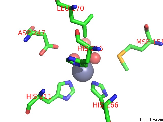
Mono view
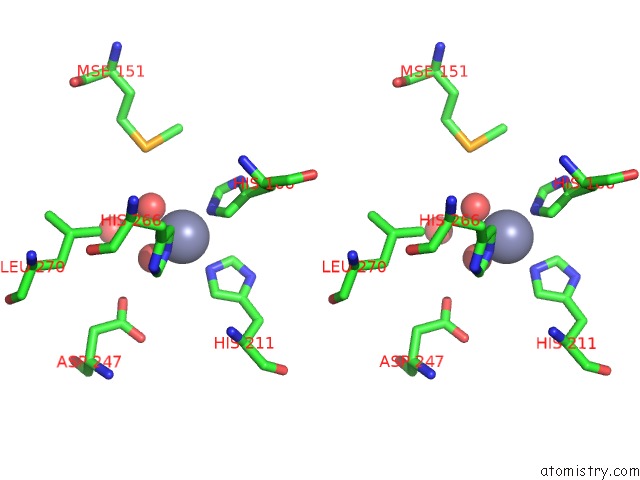
Stereo pair view
Mono view
Stereo pair view
A full contact list of Zinc with other atoms in the Zn binding
site number 1 of Crystal Structure of E.Coli Pdxa within 5.0Å range:
|
Zinc binding site 2 out of 2 in 1ptm
Go back to
Zinc binding site 2 out
of 2 in the Crystal Structure of E.Coli Pdxa
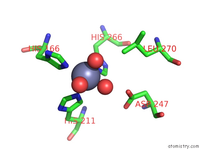
Mono view
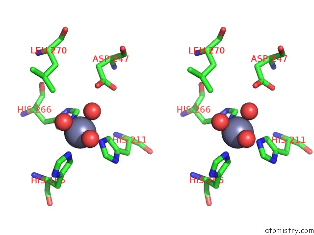
Stereo pair view
Mono view
Stereo pair view
A full contact list of Zinc with other atoms in the Zn binding
site number 2 of Crystal Structure of E.Coli Pdxa within 5.0Å range:
|
Reference:
J.Sivaraman,
Y.Li,
J.Banks,
D.E.Cane,
A.Matte,
M.Cygler.
Crystal Structure of Escherichia Coli Pdxa, An Enzyme Involved in the Pyridoxal Phosphate Biosynthesis Pathway J.Biol.Chem. V. 278 43682 2003.
ISSN: ISSN 0021-9258
PubMed: 12896974
DOI: 10.1074/JBC.M306344200
Page generated: Wed Oct 16 17:54:20 2024
ISSN: ISSN 0021-9258
PubMed: 12896974
DOI: 10.1074/JBC.M306344200
Last articles
Zn in 9J0NZn in 9J0O
Zn in 9J0P
Zn in 9FJX
Zn in 9EKB
Zn in 9C0F
Zn in 9CAH
Zn in 9CH0
Zn in 9CH3
Zn in 9CH1