Zinc »
PDB 1peg-1pv9 »
1pok »
Zinc in PDB 1pok: Crystal Structure of Isoaspartyl Dipeptidase
Protein crystallography data
The structure of Crystal Structure of Isoaspartyl Dipeptidase, PDB code: 1pok
was solved by
D.Jozic,
J.T.Kaiser,
R.Huber,
W.Bode,
K.Maskos,
with X-Ray Crystallography technique. A brief refinement statistics is given in the table below:
| Resolution Low / High (Å) | 20.00 / 2.70 |
| Space group | P 4 21 2 |
| Cell size a, b, c (Å), α, β, γ (°) | 117.475, 117.475, 137.955, 90.00, 90.00, 90.00 |
| R / Rfree (%) | 20.9 / 26.6 |
Zinc Binding Sites:
The binding sites of Zinc atom in the Crystal Structure of Isoaspartyl Dipeptidase
(pdb code 1pok). This binding sites where shown within
5.0 Angstroms radius around Zinc atom.
In total 4 binding sites of Zinc where determined in the Crystal Structure of Isoaspartyl Dipeptidase, PDB code: 1pok:
Jump to Zinc binding site number: 1; 2; 3; 4;
In total 4 binding sites of Zinc where determined in the Crystal Structure of Isoaspartyl Dipeptidase, PDB code: 1pok:
Jump to Zinc binding site number: 1; 2; 3; 4;
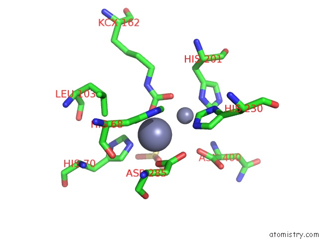
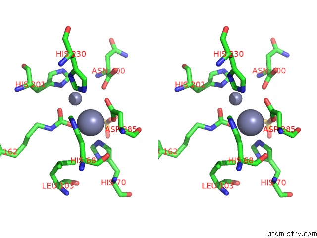
Zinc binding site 1 out of 4 in 1pok
Go back to
Zinc binding site 1 out
of 4 in the Crystal Structure of Isoaspartyl Dipeptidase
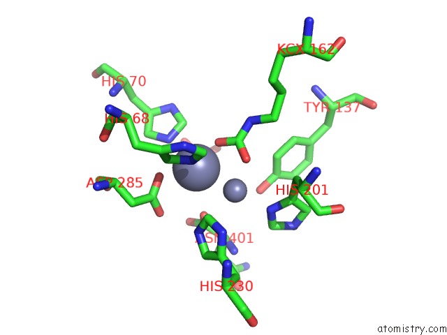
Mono view
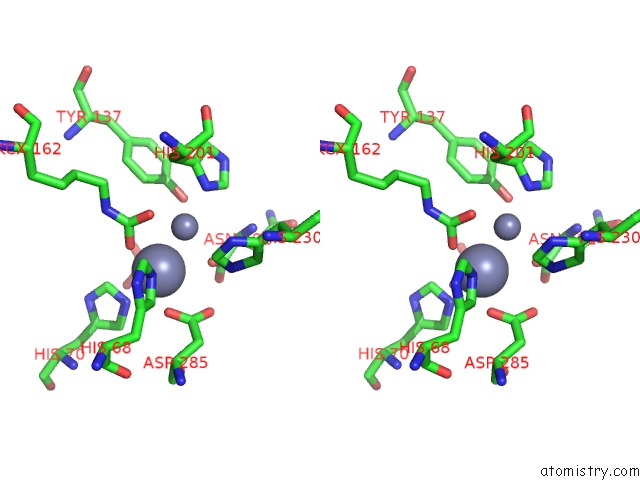
Stereo pair view
Mono view
Stereo pair view
A full contact list of Zinc with other atoms in the Zn binding
site number 1 of Crystal Structure of Isoaspartyl Dipeptidase within 5.0Å range:
|
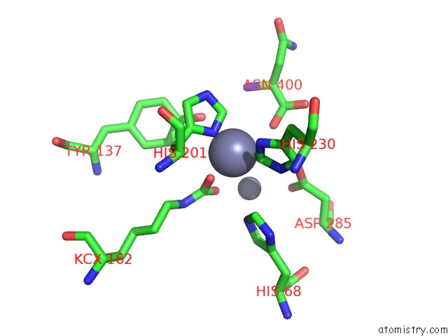
Zinc binding site 2 out of 4 in 1pok
Go back to
Zinc binding site 2 out
of 4 in the Crystal Structure of Isoaspartyl Dipeptidase
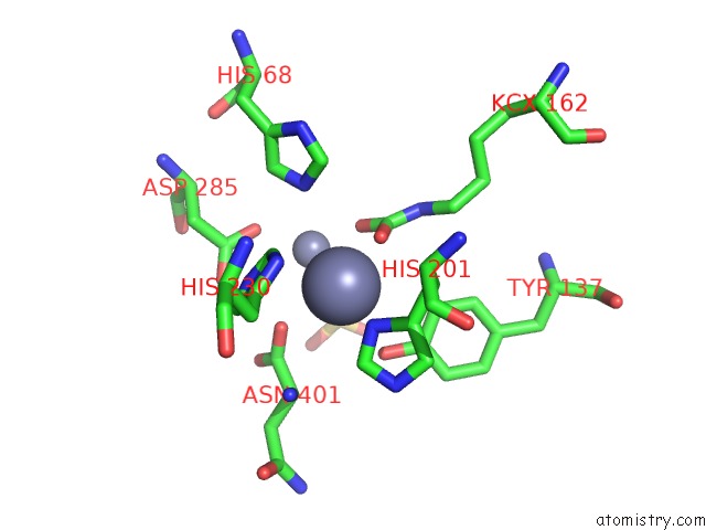
Mono view
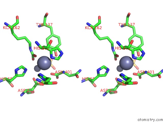
Stereo pair view
Mono view
Stereo pair view
A full contact list of Zinc with other atoms in the Zn binding
site number 2 of Crystal Structure of Isoaspartyl Dipeptidase within 5.0Å range:
|
Zinc binding site 3 out of 4 in 1pok
Go back to
Zinc binding site 3 out
of 4 in the Crystal Structure of Isoaspartyl Dipeptidase
Mono view
Stereo pair view
Mono view
Stereo pair view
A full contact list of Zinc with other atoms in the Zn binding
site number 3 of Crystal Structure of Isoaspartyl Dipeptidase within 5.0Å range:
|
Zinc binding site 4 out of 4 in 1pok
Go back to
Zinc binding site 4 out
of 4 in the Crystal Structure of Isoaspartyl Dipeptidase
Mono view

Stereo pair view
Mono view
Stereo pair view
A full contact list of Zinc with other atoms in the Zn binding
site number 4 of Crystal Structure of Isoaspartyl Dipeptidase within 5.0Å range:
|
Reference:
D.Jozic,
J.T.Kaiser,
R.Huber,
W.Bode,
K.Maskos.
X-Ray Structure of Isoaspartyl Dipeptidase From E.Coli: A Dinuclear Zinc Peptidase Evolved From Amidohydrolases. J.Mol.Biol. V. 332 243 2003.
ISSN: ISSN 0022-2836
PubMed: 12946361
DOI: 10.1016/S0022-2836(03)00845-3
Page generated: Wed Oct 16 17:52:05 2024
ISSN: ISSN 0022-2836
PubMed: 12946361
DOI: 10.1016/S0022-2836(03)00845-3
Last articles
Zn in 9J0NZn in 9J0O
Zn in 9J0P
Zn in 9FJX
Zn in 9EKB
Zn in 9C0F
Zn in 9CAH
Zn in 9CH0
Zn in 9CH3
Zn in 9CH1