Zinc »
PDB 1peg-1pv9 »
1pi1 »
Zinc in PDB 1pi1: Crystal Structure of A Human MOB1 Protein; Toward Understanding Mob-Regulated Cell Cycle Pathways.
Protein crystallography data
The structure of Crystal Structure of A Human MOB1 Protein; Toward Understanding Mob-Regulated Cell Cycle Pathways., PDB code: 1pi1
was solved by
E.S.Stavridi,
K.G.Harris,
Y.Huyen,
J.Bothos,
P.M.Voewerd,
S.E.Stayrook,
P.D.Jeffrey,
N.P.Pavletich,
F.C.Luca,
with X-Ray Crystallography technique. A brief refinement statistics is given in the table below:
| Resolution Low / High (Å) | 25.00 / 2.00 |
| Space group | P 32 2 1 |
| Cell size a, b, c (Å), α, β, γ (°) | 67.676, 67.676, 95.595, 90.00, 90.00, 120.00 |
| R / Rfree (%) | 20.5 / 24.2 |
Zinc Binding Sites:
The binding sites of Zinc atom in the Crystal Structure of A Human MOB1 Protein; Toward Understanding Mob-Regulated Cell Cycle Pathways.
(pdb code 1pi1). This binding sites where shown within
5.0 Angstroms radius around Zinc atom.
In total only one binding site of Zinc was determined in the Crystal Structure of A Human MOB1 Protein; Toward Understanding Mob-Regulated Cell Cycle Pathways., PDB code: 1pi1:
In total only one binding site of Zinc was determined in the Crystal Structure of A Human MOB1 Protein; Toward Understanding Mob-Regulated Cell Cycle Pathways., PDB code: 1pi1:
Zinc binding site 1 out of 1 in 1pi1
Go back to
Zinc binding site 1 out
of 1 in the Crystal Structure of A Human MOB1 Protein; Toward Understanding Mob-Regulated Cell Cycle Pathways.
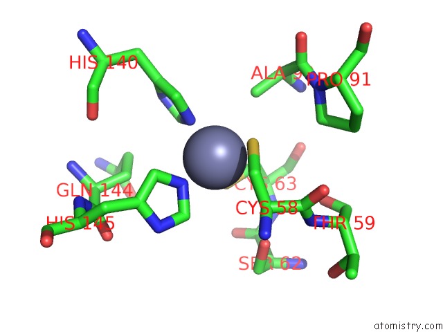
Mono view
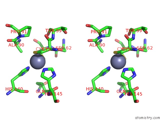
Stereo pair view
Mono view
Stereo pair view
A full contact list of Zinc with other atoms in the Zn binding
site number 1 of Crystal Structure of A Human MOB1 Protein; Toward Understanding Mob-Regulated Cell Cycle Pathways. within 5.0Å range:
|
Reference:
E.S.Stavridi,
K.G.Harris,
Y.Huyen,
J.Bothos,
P.M.Verwoerd,
S.E.Stayrook,
N.P.Pavletich,
P.D.Jeffrey,
F.C.Luca.
Crystal Structure of A Human MOB1 Protein. Toward Understanding Mob-Regulated Cell Cycle Pathways. Structure V. 11 1163 2003.
ISSN: ISSN 0969-2126
PubMed: 12962634
DOI: 10.1016/S0969-2126(03)00182-5
Page generated: Wed Oct 16 17:49:44 2024
ISSN: ISSN 0969-2126
PubMed: 12962634
DOI: 10.1016/S0969-2126(03)00182-5
Last articles
Zn in 9J0NZn in 9J0O
Zn in 9J0P
Zn in 9FJX
Zn in 9EKB
Zn in 9C0F
Zn in 9CAH
Zn in 9CH0
Zn in 9CH3
Zn in 9CH1