Zinc »
PDB 1peg-1pv9 »
1pfw »
Zinc in PDB 1pfw: Methionyl-Trna Synthetase From Escherichia Coli Complexed with Trifluoromethionine
Enzymatic activity of Methionyl-Trna Synthetase From Escherichia Coli Complexed with Trifluoromethionine
All present enzymatic activity of Methionyl-Trna Synthetase From Escherichia Coli Complexed with Trifluoromethionine:
6.1.1.10;
6.1.1.10;
Protein crystallography data
The structure of Methionyl-Trna Synthetase From Escherichia Coli Complexed with Trifluoromethionine, PDB code: 1pfw
was solved by
T.Crepin,
E.Schmitt,
Y.Mechulam,
P.B.Sampson,
M.D.Vaughan,
J.F.Honek,
S.Blanquet,
with X-Ray Crystallography technique. A brief refinement statistics is given in the table below:
| Resolution Low / High (Å) | 45.00 / 1.78 |
| Space group | P 1 21 1 |
| Cell size a, b, c (Å), α, β, γ (°) | 78.490, 45.288, 86.274, 90.00, 107.37, 90.00 |
| R / Rfree (%) | 18.4 / 20.9 |
Other elements in 1pfw:
The structure of Methionyl-Trna Synthetase From Escherichia Coli Complexed with Trifluoromethionine also contains other interesting chemical elements:
| Fluorine | (F) | 3 atoms |
Zinc Binding Sites:
The binding sites of Zinc atom in the Methionyl-Trna Synthetase From Escherichia Coli Complexed with Trifluoromethionine
(pdb code 1pfw). This binding sites where shown within
5.0 Angstroms radius around Zinc atom.
In total only one binding site of Zinc was determined in the Methionyl-Trna Synthetase From Escherichia Coli Complexed with Trifluoromethionine, PDB code: 1pfw:
In total only one binding site of Zinc was determined in the Methionyl-Trna Synthetase From Escherichia Coli Complexed with Trifluoromethionine, PDB code: 1pfw:
Zinc binding site 1 out of 1 in 1pfw
Go back to
Zinc binding site 1 out
of 1 in the Methionyl-Trna Synthetase From Escherichia Coli Complexed with Trifluoromethionine
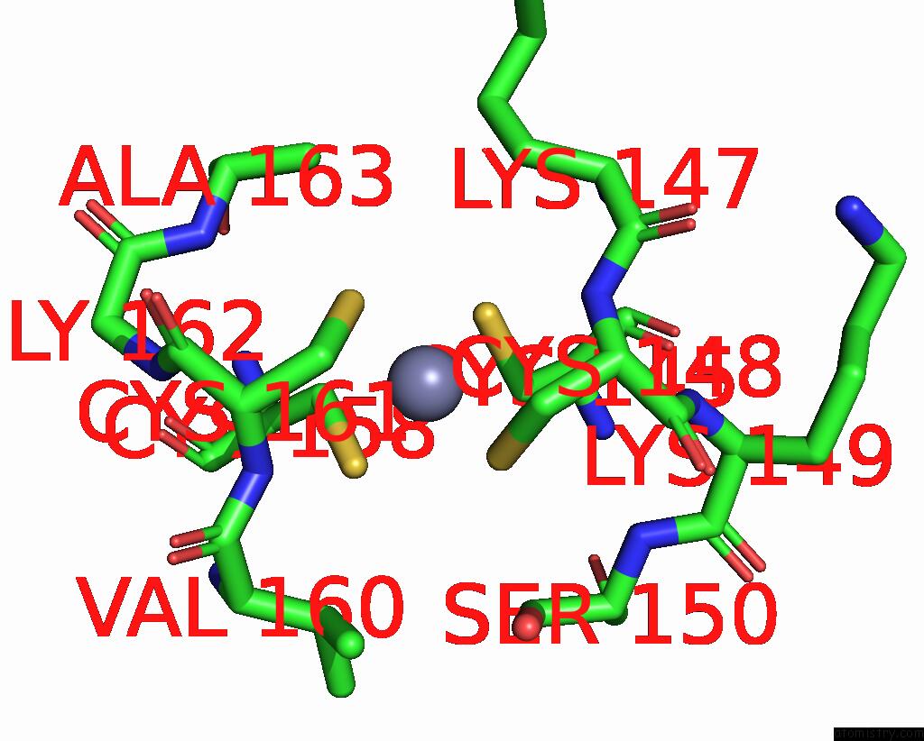
Mono view
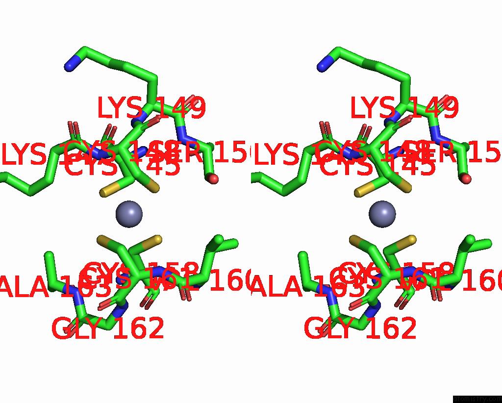
Stereo pair view
Mono view
Stereo pair view
A full contact list of Zinc with other atoms in the Zn binding
site number 1 of Methionyl-Trna Synthetase From Escherichia Coli Complexed with Trifluoromethionine within 5.0Å range:
|
Reference:
T.Crepin,
E.Schmitt,
Y.Mechulam,
P.B.Sampson,
M.D.Vaughan,
J.F.Honek,
S.Blanquet.
Use of Analogues of Methionine and Methionyl Adenylate to Sample Conformational Changes During Catalysis in Escherichia Coli Methionyl-Trna Synthetase. J.Mol.Biol. V. 332 59 2003.
ISSN: ISSN 0022-2836
PubMed: 12946347
DOI: 10.1016/S0022-2836(03)00917-3
Page generated: Wed Oct 16 17:49:16 2024
ISSN: ISSN 0022-2836
PubMed: 12946347
DOI: 10.1016/S0022-2836(03)00917-3
Last articles
Zn in 9J0NZn in 9J0O
Zn in 9J0P
Zn in 9FJX
Zn in 9EKB
Zn in 9C0F
Zn in 9CAH
Zn in 9CH0
Zn in 9CH3
Zn in 9CH1