Zinc »
PDB 1oj7-1p1r »
1p0d »
Zinc in PDB 1p0d: Crystal Structure of Zymomonas Mobilis Trna-Guanine Transglycosylase (Tgt) Crystallised at pH 5.5
Enzymatic activity of Crystal Structure of Zymomonas Mobilis Trna-Guanine Transglycosylase (Tgt) Crystallised at pH 5.5
All present enzymatic activity of Crystal Structure of Zymomonas Mobilis Trna-Guanine Transglycosylase (Tgt) Crystallised at pH 5.5:
2.4.2.29;
2.4.2.29;
Protein crystallography data
The structure of Crystal Structure of Zymomonas Mobilis Trna-Guanine Transglycosylase (Tgt) Crystallised at pH 5.5, PDB code: 1p0d
was solved by
R.Brenk,
M.T.Stubbs,
A.Heine,
K.Reuter,
G.Klebe,
with X-Ray Crystallography technique. A brief refinement statistics is given in the table below:
| Resolution Low / High (Å) | 32.69 / 1.90 |
| Space group | C 1 2 1 |
| Cell size a, b, c (Å), α, β, γ (°) | 90.380, 65.370, 70.570, 90.00, 96.30, 90.00 |
| R / Rfree (%) | 17.2 / 20.9 |
Zinc Binding Sites:
The binding sites of Zinc atom in the Crystal Structure of Zymomonas Mobilis Trna-Guanine Transglycosylase (Tgt) Crystallised at pH 5.5
(pdb code 1p0d). This binding sites where shown within
5.0 Angstroms radius around Zinc atom.
In total only one binding site of Zinc was determined in the Crystal Structure of Zymomonas Mobilis Trna-Guanine Transglycosylase (Tgt) Crystallised at pH 5.5, PDB code: 1p0d:
In total only one binding site of Zinc was determined in the Crystal Structure of Zymomonas Mobilis Trna-Guanine Transglycosylase (Tgt) Crystallised at pH 5.5, PDB code: 1p0d:
Zinc binding site 1 out of 1 in 1p0d
Go back to
Zinc binding site 1 out
of 1 in the Crystal Structure of Zymomonas Mobilis Trna-Guanine Transglycosylase (Tgt) Crystallised at pH 5.5
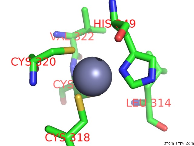
Mono view
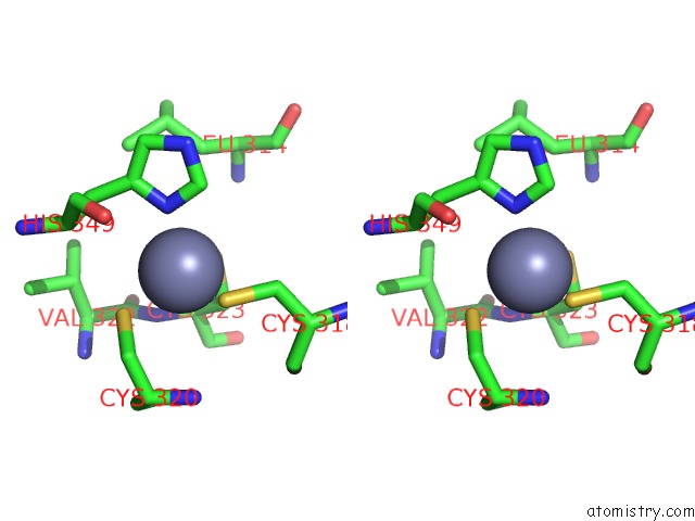
Stereo pair view
Mono view
Stereo pair view
A full contact list of Zinc with other atoms in the Zn binding
site number 1 of Crystal Structure of Zymomonas Mobilis Trna-Guanine Transglycosylase (Tgt) Crystallised at pH 5.5 within 5.0Å range:
|
Reference:
R.Brenk,
M.T.Stubbs,
A.Heine,
K.Reuter,
G.Klebe.
Flexible Adaptations in the Structure of the Trna-Modifying Enzyme Trna-Guanine Transglycosylase and Their Implications For Substrate Selectivity, Reaction Mechanism and Structure-Based Drug Design Chembiochem V. 4 1066 2003.
ISSN: ISSN 1439-4227
PubMed: 14523925
DOI: 10.1002/CBIC.200300644
Page generated: Tue Aug 19 22:15:24 2025
ISSN: ISSN 1439-4227
PubMed: 14523925
DOI: 10.1002/CBIC.200300644
Last articles
Zn in 1ZY1Zn in 1ZY7
Zn in 1ZY0
Zn in 1ZXZ
Zn in 1ZXV
Zn in 1ZXC
Zn in 1ZWJ
Zn in 1ZX1
Zn in 1ZTQ
Zn in 1ZW8