Zinc »
PDB 1oj7-1p1r »
1ozj »
Zinc in PDB 1ozj: Crystal Structure of SMAD3-MH1 Bound to Dna at 2.4 A Resolution
Protein crystallography data
The structure of Crystal Structure of SMAD3-MH1 Bound to Dna at 2.4 A Resolution, PDB code: 1ozj
was solved by
J.Chai,
J.-W.Wu,
N.Yan,
J.Massague,
N.P.Pavletich,
Y.Shi,
with X-Ray Crystallography technique. A brief refinement statistics is given in the table below:
| Resolution Low / High (Å) | 15.00 / 2.40 |
| Space group | P 1 21 1 |
| Cell size a, b, c (Å), α, β, γ (°) | 45.600, 60.400, 71.600, 90.00, 102.00, 90.00 |
| R / Rfree (%) | 21 / 27 |
Zinc Binding Sites:
The binding sites of Zinc atom in the Crystal Structure of SMAD3-MH1 Bound to Dna at 2.4 A Resolution
(pdb code 1ozj). This binding sites where shown within
5.0 Angstroms radius around Zinc atom.
In total 2 binding sites of Zinc where determined in the Crystal Structure of SMAD3-MH1 Bound to Dna at 2.4 A Resolution, PDB code: 1ozj:
Jump to Zinc binding site number: 1; 2;
In total 2 binding sites of Zinc where determined in the Crystal Structure of SMAD3-MH1 Bound to Dna at 2.4 A Resolution, PDB code: 1ozj:
Jump to Zinc binding site number: 1; 2;
Zinc binding site 1 out of 2 in 1ozj
Go back to
Zinc binding site 1 out
of 2 in the Crystal Structure of SMAD3-MH1 Bound to Dna at 2.4 A Resolution
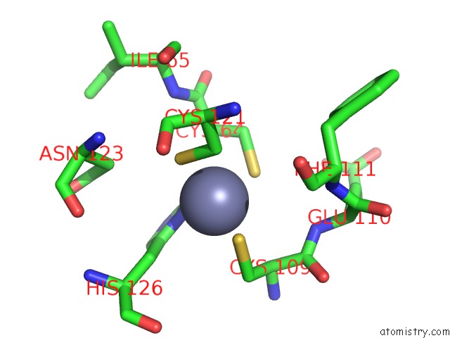
Mono view
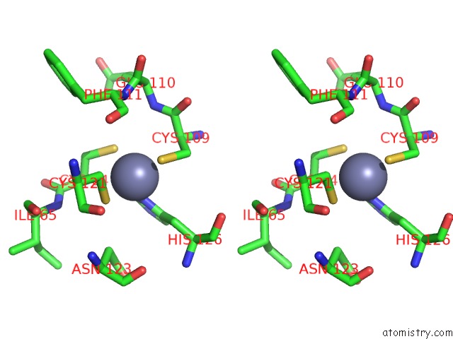
Stereo pair view
Mono view
Stereo pair view
A full contact list of Zinc with other atoms in the Zn binding
site number 1 of Crystal Structure of SMAD3-MH1 Bound to Dna at 2.4 A Resolution within 5.0Å range:
|
Zinc binding site 2 out of 2 in 1ozj
Go back to
Zinc binding site 2 out
of 2 in the Crystal Structure of SMAD3-MH1 Bound to Dna at 2.4 A Resolution
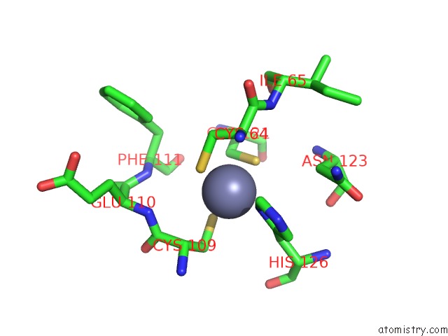
Mono view
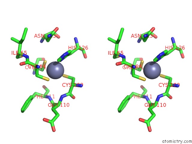
Stereo pair view
Mono view
Stereo pair view
A full contact list of Zinc with other atoms in the Zn binding
site number 2 of Crystal Structure of SMAD3-MH1 Bound to Dna at 2.4 A Resolution within 5.0Å range:
|
Reference:
J.Chai,
J.-W.Wu,
N.Yan,
J.Massague,
N.P.Pavletich,
Y.Shi.
Features of A SMAD3 MH1-Dna Complex. Roles of Water and Zinc in Dna Binding. J.Biol.Chem. V. 278 20327 2003.
ISSN: ISSN 0021-9258
PubMed: 12686552
DOI: 10.1074/JBC.C300134200
Page generated: Tue Aug 19 22:14:38 2025
ISSN: ISSN 0021-9258
PubMed: 12686552
DOI: 10.1074/JBC.C300134200
Last articles
Zn in 1ZQTZn in 1ZNS
Zn in 1ZNB
Zn in 1ZNJ
Zn in 1ZNM
Zn in 1ZNI
Zn in 1ZNC
Zn in 1ZNF
Zn in 1ZME
Zn in 1ZLH