Zinc »
PDB 1oj7-1p1r »
1oxq »
Zinc in PDB 1oxq: Structure and Function Analysis of Peptide Antagonists of Melanoma Inhibitor of Apoptosis (Ml-Iap)
Protein crystallography data
The structure of Structure and Function Analysis of Peptide Antagonists of Melanoma Inhibitor of Apoptosis (Ml-Iap), PDB code: 1oxq
was solved by
M.C.Franklin,
S.Kadkhodayan,
H.Ackerly,
D.Alexandru,
M.D.Distefano,
L.O.Elliott,
J.A.Flygare,
D.Vucic,
K.Deshayes,
W.J.Fairbrother,
with X-Ray Crystallography technique. A brief refinement statistics is given in the table below:
| Resolution Low / High (Å) | 20.00 / 2.30 |
| Space group | P 32 |
| Cell size a, b, c (Å), α, β, γ (°) | 83.189, 83.189, 93.612, 90.00, 90.00, 120.00 |
| R / Rfree (%) | 16.1 / 21.8 |
Zinc Binding Sites:
The binding sites of Zinc atom in the Structure and Function Analysis of Peptide Antagonists of Melanoma Inhibitor of Apoptosis (Ml-Iap)
(pdb code 1oxq). This binding sites where shown within
5.0 Angstroms radius around Zinc atom.
In total 5 binding sites of Zinc where determined in the Structure and Function Analysis of Peptide Antagonists of Melanoma Inhibitor of Apoptosis (Ml-Iap), PDB code: 1oxq:
Jump to Zinc binding site number: 1; 2; 3; 4; 5;
In total 5 binding sites of Zinc where determined in the Structure and Function Analysis of Peptide Antagonists of Melanoma Inhibitor of Apoptosis (Ml-Iap), PDB code: 1oxq:
Jump to Zinc binding site number: 1; 2; 3; 4; 5;
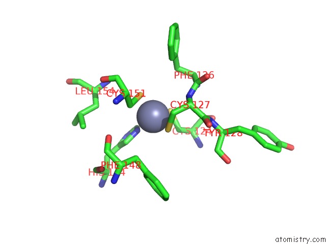
Zinc binding site 1 out of 5 in 1oxq
Go back to
Zinc binding site 1 out
of 5 in the Structure and Function Analysis of Peptide Antagonists of Melanoma Inhibitor of Apoptosis (Ml-Iap)

Mono view

Stereo pair view
Mono view
Stereo pair view
A full contact list of Zinc with other atoms in the Zn binding
site number 1 of Structure and Function Analysis of Peptide Antagonists of Melanoma Inhibitor of Apoptosis (Ml-Iap) within 5.0Å range:
|
Zinc binding site 2 out of 5 in 1oxq
Go back to
Zinc binding site 2 out
of 5 in the Structure and Function Analysis of Peptide Antagonists of Melanoma Inhibitor of Apoptosis (Ml-Iap)
Mono view

Stereo pair view
Mono view
Stereo pair view
A full contact list of Zinc with other atoms in the Zn binding
site number 2 of Structure and Function Analysis of Peptide Antagonists of Melanoma Inhibitor of Apoptosis (Ml-Iap) within 5.0Å range:
|
Zinc binding site 3 out of 5 in 1oxq
Go back to
Zinc binding site 3 out
of 5 in the Structure and Function Analysis of Peptide Antagonists of Melanoma Inhibitor of Apoptosis (Ml-Iap)

Mono view

Stereo pair view
Mono view
Stereo pair view
A full contact list of Zinc with other atoms in the Zn binding
site number 3 of Structure and Function Analysis of Peptide Antagonists of Melanoma Inhibitor of Apoptosis (Ml-Iap) within 5.0Å range:
|
Zinc binding site 4 out of 5 in 1oxq
Go back to
Zinc binding site 4 out
of 5 in the Structure and Function Analysis of Peptide Antagonists of Melanoma Inhibitor of Apoptosis (Ml-Iap)

Mono view

Stereo pair view
Mono view
Stereo pair view
A full contact list of Zinc with other atoms in the Zn binding
site number 4 of Structure and Function Analysis of Peptide Antagonists of Melanoma Inhibitor of Apoptosis (Ml-Iap) within 5.0Å range:
|
Zinc binding site 5 out of 5 in 1oxq
Go back to
Zinc binding site 5 out
of 5 in the Structure and Function Analysis of Peptide Antagonists of Melanoma Inhibitor of Apoptosis (Ml-Iap)

Mono view

Stereo pair view
Mono view
Stereo pair view
A full contact list of Zinc with other atoms in the Zn binding
site number 5 of Structure and Function Analysis of Peptide Antagonists of Melanoma Inhibitor of Apoptosis (Ml-Iap) within 5.0Å range:
|
Reference:
M.C.Franklin,
S.Kadkhodayan,
H.Ackerly,
D.Alexandru,
M.D.Distefano,
L.O.Elliott,
J.A.Flygare,
G.Mausisa,
D.C.Okawa,
D.Ong,
D.Vucic,
K.Deshayes,
W.J.Fairbrother.
Structure and Function Analysis of Peptide Antagonists of Melanoma Inhibitor of Apoptosis (Ml-Iap) Biochemistry V. 42 8223 2003.
ISSN: ISSN 0006-2960
PubMed: 12846571
DOI: 10.1021/BI034227T
Page generated: Wed Oct 16 17:36:29 2024
ISSN: ISSN 0006-2960
PubMed: 12846571
DOI: 10.1021/BI034227T
Last articles
Zn in 9J0NZn in 9J0O
Zn in 9J0P
Zn in 9FJX
Zn in 9EKB
Zn in 9C0F
Zn in 9CAH
Zn in 9CH0
Zn in 9CH3
Zn in 9CH1