Zinc in PDB 1ovx: uc(Nmr) Structure of the E. Coli Clpx Chaperone Zinc Binding Domain Dimer
Zinc Binding Sites:
The binding sites of Zinc atom in the uc(Nmr) Structure of the E. Coli Clpx Chaperone Zinc Binding Domain Dimer
(pdb code 1ovx). This binding sites where shown within
5.0 Angstroms radius around Zinc atom.
In total 2 binding sites of Zinc where determined in the
uc(Nmr) Structure of the E. Coli Clpx Chaperone Zinc Binding Domain Dimer, PDB code: 1ovx:
Jump to Zinc binding site number:
1;
2;
Zinc binding site 1 out
of 2 in 1ovx
Go back to
Zinc Binding Sites List in 1ovx
Zinc binding site 1 out
of 2 in the uc(Nmr) Structure of the E. Coli Clpx Chaperone Zinc Binding Domain Dimer
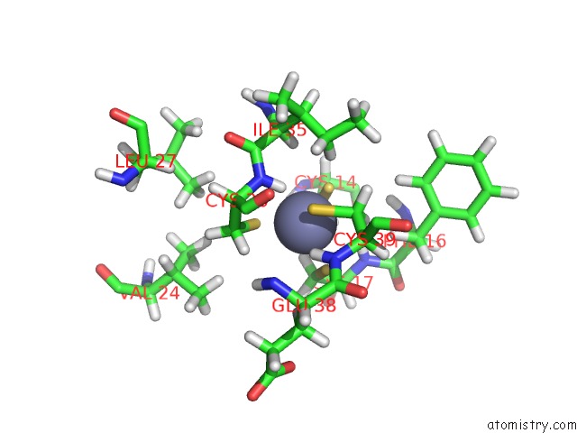
Mono view
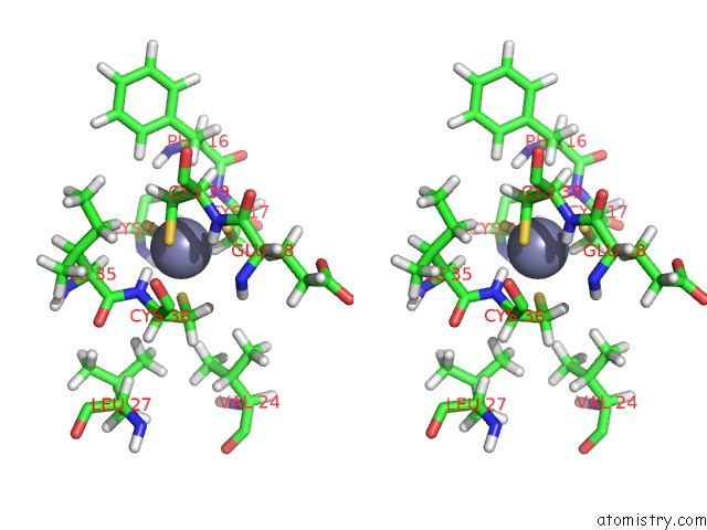
Stereo pair view
|
A full contact list of Zinc with other atoms in the Zn binding
site number 1 of uc(Nmr) Structure of the E. Coli Clpx Chaperone Zinc Binding Domain Dimer within 5.0Å range:
probe
|
atom
|
residue
|
distance (Å)
|
B
|
Occ
|
A:Zn61
b:0.0
occ:1.00
|
H
|
A:CYS36
|
2.1
|
0.0
|
1.0
|
SG
|
A:CYS39
|
2.3
|
0.0
|
1.0
|
SG
|
A:CYS36
|
2.3
|
0.0
|
1.0
|
SG
|
A:CYS14
|
2.3
|
0.0
|
1.0
|
SG
|
A:CYS17
|
2.3
|
0.0
|
1.0
|
HB3
|
A:CYS39
|
2.8
|
0.0
|
1.0
|
HB2
|
A:CYS17
|
3.0
|
0.0
|
1.0
|
N
|
A:CYS36
|
3.0
|
0.0
|
1.0
|
HB2
|
A:CYS14
|
3.1
|
0.0
|
1.0
|
CB
|
A:CYS39
|
3.1
|
0.0
|
1.0
|
CB
|
A:CYS14
|
3.2
|
0.0
|
1.0
|
CB
|
A:CYS17
|
3.2
|
0.0
|
1.0
|
CB
|
A:CYS36
|
3.3
|
0.0
|
1.0
|
HB2
|
A:CYS36
|
3.3
|
0.0
|
1.0
|
H
|
A:CYS39
|
3.3
|
0.0
|
1.0
|
HG13
|
A:ILE35
|
3.3
|
0.0
|
1.0
|
H
|
A:CYS17
|
3.4
|
0.0
|
1.0
|
HB3
|
A:CYS14
|
3.4
|
0.0
|
1.0
|
HA
|
A:ILE35
|
3.5
|
0.0
|
1.0
|
CA
|
A:CYS36
|
3.6
|
0.0
|
1.0
|
HB2
|
A:CYS39
|
3.9
|
0.0
|
1.0
|
HB3
|
A:CYS17
|
3.9
|
0.0
|
1.0
|
N
|
A:CYS39
|
4.0
|
0.0
|
1.0
|
C
|
A:ILE35
|
4.0
|
0.0
|
1.0
|
HB2
|
A:PHE16
|
4.1
|
0.0
|
1.0
|
N
|
A:CYS17
|
4.1
|
0.0
|
1.0
|
CA
|
A:CYS39
|
4.1
|
0.0
|
1.0
|
C
|
A:CYS36
|
4.2
|
0.0
|
1.0
|
CG1
|
A:ILE35
|
4.2
|
0.0
|
1.0
|
HG12
|
A:ILE35
|
4.2
|
0.0
|
1.0
|
HB3
|
A:CYS36
|
4.3
|
0.0
|
1.0
|
CA
|
A:ILE35
|
4.3
|
0.0
|
1.0
|
O
|
A:CYS36
|
4.3
|
0.0
|
1.0
|
CA
|
A:CYS17
|
4.3
|
0.0
|
1.0
|
HB2
|
A:GLU38
|
4.5
|
0.0
|
1.0
|
HA
|
A:CYS39
|
4.5
|
0.0
|
1.0
|
CA
|
A:CYS14
|
4.6
|
0.0
|
1.0
|
HA
|
A:CYS36
|
4.6
|
0.0
|
1.0
|
HG13
|
A:VAL24
|
4.6
|
0.0
|
1.0
|
HD2
|
A:PHE16
|
4.6
|
0.0
|
1.0
|
HA
|
A:CYS14
|
4.7
|
0.0
|
1.0
|
HD23
|
A:LEU27
|
4.8
|
0.0
|
1.0
|
HG22
|
A:VAL24
|
4.8
|
0.0
|
1.0
|
H
|
A:PHE16
|
4.9
|
0.0
|
1.0
|
CB
|
A:ILE35
|
4.9
|
0.0
|
1.0
|
H
|
A:GLU38
|
4.9
|
0.0
|
1.0
|
HA
|
A:CYS17
|
5.0
|
0.0
|
1.0
|
|
Zinc binding site 2 out
of 2 in 1ovx
Go back to
Zinc Binding Sites List in 1ovx
Zinc binding site 2 out
of 2 in the uc(Nmr) Structure of the E. Coli Clpx Chaperone Zinc Binding Domain Dimer
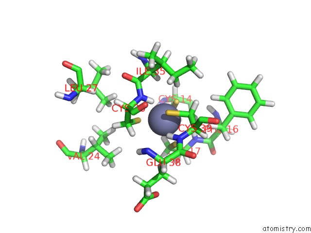
Mono view
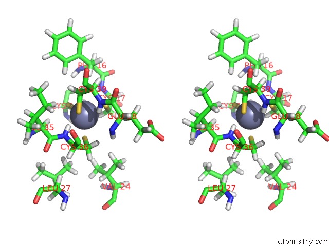
Stereo pair view
|
A full contact list of Zinc with other atoms in the Zn binding
site number 2 of uc(Nmr) Structure of the E. Coli Clpx Chaperone Zinc Binding Domain Dimer within 5.0Å range:
probe
|
atom
|
residue
|
distance (Å)
|
B
|
Occ
|
B:Zn61
b:0.0
occ:1.00
|
H
|
B:CYS36
|
2.1
|
0.0
|
1.0
|
SG
|
B:CYS39
|
2.3
|
0.0
|
1.0
|
SG
|
B:CYS36
|
2.3
|
0.0
|
1.0
|
SG
|
B:CYS14
|
2.3
|
0.0
|
1.0
|
SG
|
B:CYS17
|
2.3
|
0.0
|
1.0
|
HB3
|
B:CYS39
|
2.8
|
0.0
|
1.0
|
HB2
|
B:CYS17
|
3.0
|
0.0
|
1.0
|
N
|
B:CYS36
|
3.0
|
0.0
|
1.0
|
HB2
|
B:CYS14
|
3.1
|
0.0
|
1.0
|
CB
|
B:CYS39
|
3.1
|
0.0
|
1.0
|
CB
|
B:CYS14
|
3.2
|
0.0
|
1.0
|
CB
|
B:CYS17
|
3.2
|
0.0
|
1.0
|
CB
|
B:CYS36
|
3.3
|
0.0
|
1.0
|
HB2
|
B:CYS36
|
3.3
|
0.0
|
1.0
|
H
|
B:CYS39
|
3.3
|
0.0
|
1.0
|
HG13
|
B:ILE35
|
3.3
|
0.0
|
1.0
|
H
|
B:CYS17
|
3.4
|
0.0
|
1.0
|
HB3
|
B:CYS14
|
3.4
|
0.0
|
1.0
|
HA
|
B:ILE35
|
3.6
|
0.0
|
1.0
|
CA
|
B:CYS36
|
3.6
|
0.0
|
1.0
|
HB2
|
B:CYS39
|
3.9
|
0.0
|
1.0
|
HB3
|
B:CYS17
|
3.9
|
0.0
|
1.0
|
N
|
B:CYS39
|
4.0
|
0.0
|
1.0
|
HB2
|
B:PHE16
|
4.0
|
0.0
|
1.0
|
C
|
B:ILE35
|
4.0
|
0.0
|
1.0
|
N
|
B:CYS17
|
4.1
|
0.0
|
1.0
|
CA
|
B:CYS39
|
4.1
|
0.0
|
1.0
|
C
|
B:CYS36
|
4.2
|
0.0
|
1.0
|
CG1
|
B:ILE35
|
4.2
|
0.0
|
1.0
|
HG12
|
B:ILE35
|
4.2
|
0.0
|
1.0
|
HB3
|
B:CYS36
|
4.3
|
0.0
|
1.0
|
CA
|
B:ILE35
|
4.3
|
0.0
|
1.0
|
O
|
B:CYS36
|
4.3
|
0.0
|
1.0
|
CA
|
B:CYS17
|
4.3
|
0.0
|
1.0
|
HB2
|
B:GLU38
|
4.5
|
0.0
|
1.0
|
HA
|
B:CYS39
|
4.5
|
0.0
|
1.0
|
CA
|
B:CYS14
|
4.6
|
0.0
|
1.0
|
HD2
|
B:PHE16
|
4.6
|
0.0
|
1.0
|
HG13
|
B:VAL24
|
4.6
|
0.0
|
1.0
|
HA
|
B:CYS36
|
4.6
|
0.0
|
1.0
|
HA
|
B:CYS14
|
4.7
|
0.0
|
1.0
|
HD23
|
B:LEU27
|
4.8
|
0.0
|
1.0
|
HG22
|
B:VAL24
|
4.8
|
0.0
|
1.0
|
H
|
B:PHE16
|
4.9
|
0.0
|
1.0
|
CB
|
B:ILE35
|
4.9
|
0.0
|
1.0
|
H
|
B:GLU38
|
5.0
|
0.0
|
1.0
|
HA
|
B:CYS17
|
5.0
|
0.0
|
1.0
|
|
Reference:
L.W.Donaldson,
U.Wojtyra,
W.A.Houry.
Solution Structure of the Dimeric Zinc Binding Domain of the Chaperone Clpx. J.Biol.Chem. V. 278 48991 2003.
ISSN: ISSN 0021-9258
PubMed: 14525985
DOI: 10.1074/JBC.M307826200
Page generated: Wed Oct 16 17:33:59 2024
|