Zinc »
PDB 1oj7-1p1r »
1oj7 »
Zinc in PDB 1oj7: Structural Genomics, Unknown Function Crystal Structure of E. Coli K-12 Yqhd
Protein crystallography data
The structure of Structural Genomics, Unknown Function Crystal Structure of E. Coli K-12 Yqhd, PDB code: 1oj7
was solved by
G.Sulzenbacher,
S.Perrier,
V.Roig-Zamboni,
F.Pagot,
S.Grisel,
A.Salamoni,
C.Valencia,
C.Bignon,
R.Vincentelli,
M.Tegoni,
C.Cambillau,
with X-Ray Crystallography technique. A brief refinement statistics is given in the table below:
| Resolution Low / High (Å) | 47.00 / 2.0 |
| Space group | P 62 |
| Cell size a, b, c (Å), α, β, γ (°) | 237.925, 237.925, 66.744, 90.00, 90.00, 120.00 |
| R / Rfree (%) | 14.5 / 16.4 |
Other elements in 1oj7:
The structure of Structural Genomics, Unknown Function Crystal Structure of E. Coli K-12 Yqhd also contains other interesting chemical elements:
| Chlorine | (Cl) | 1 atom |
Zinc Binding Sites:
The binding sites of Zinc atom in the Structural Genomics, Unknown Function Crystal Structure of E. Coli K-12 Yqhd
(pdb code 1oj7). This binding sites where shown within
5.0 Angstroms radius around Zinc atom.
In total 3 binding sites of Zinc where determined in the Structural Genomics, Unknown Function Crystal Structure of E. Coli K-12 Yqhd, PDB code: 1oj7:
Jump to Zinc binding site number: 1; 2; 3;
In total 3 binding sites of Zinc where determined in the Structural Genomics, Unknown Function Crystal Structure of E. Coli K-12 Yqhd, PDB code: 1oj7:
Jump to Zinc binding site number: 1; 2; 3;
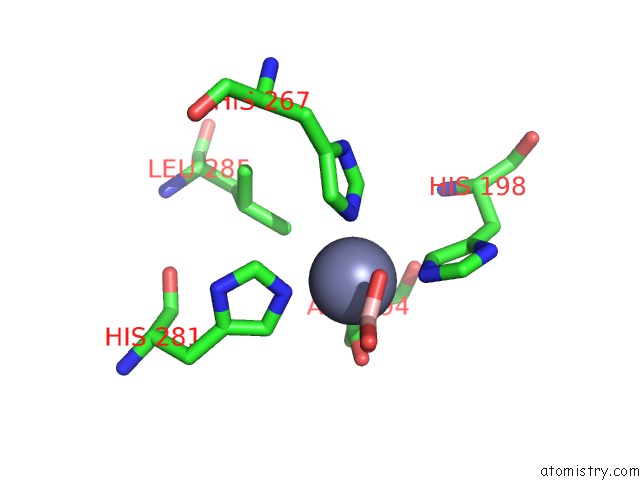
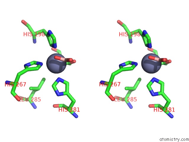
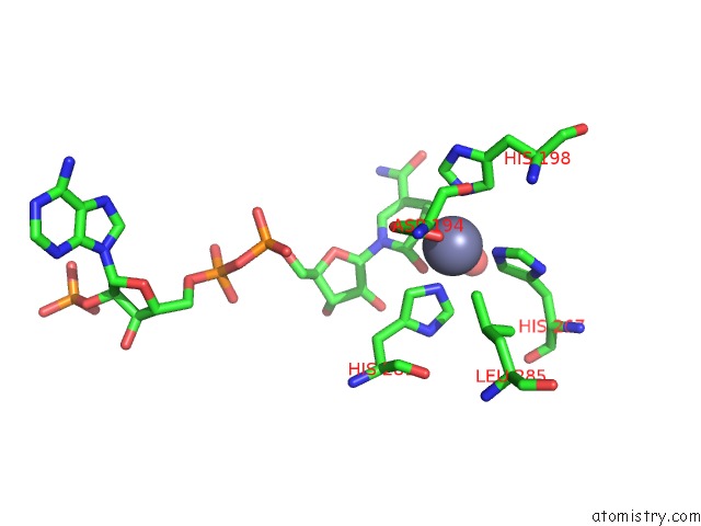
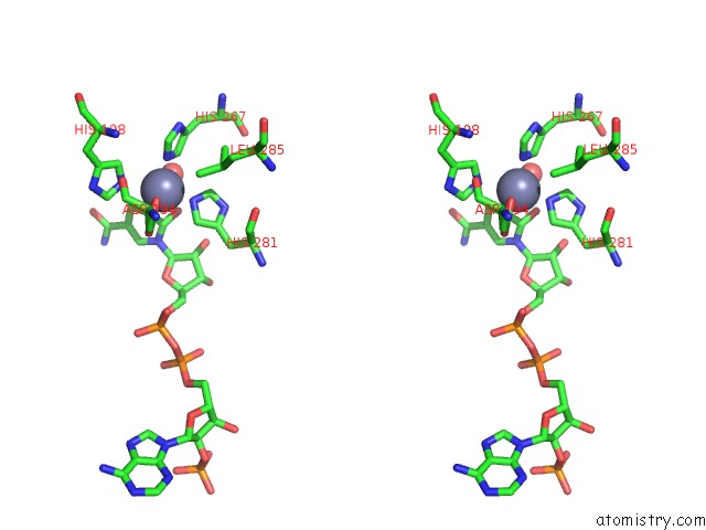
Zinc binding site 1 out of 3 in 1oj7
Go back to
Zinc binding site 1 out
of 3 in the Structural Genomics, Unknown Function Crystal Structure of E. Coli K-12 Yqhd
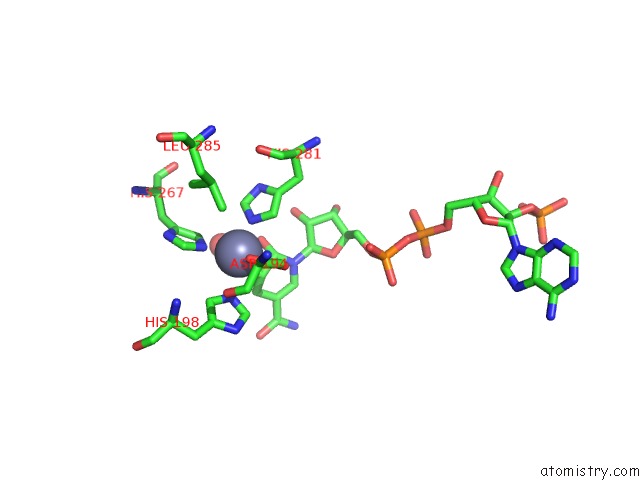
Mono view

Stereo pair view
Mono view
Stereo pair view
A full contact list of Zinc with other atoms in the Zn binding
site number 1 of Structural Genomics, Unknown Function Crystal Structure of E. Coli K-12 Yqhd within 5.0Å range:
|
Zinc binding site 2 out of 3 in 1oj7
Go back to
Zinc binding site 2 out
of 3 in the Structural Genomics, Unknown Function Crystal Structure of E. Coli K-12 Yqhd
Mono view
Stereo pair view
Mono view
Stereo pair view
A full contact list of Zinc with other atoms in the Zn binding
site number 2 of Structural Genomics, Unknown Function Crystal Structure of E. Coli K-12 Yqhd within 5.0Å range:
|
Zinc binding site 3 out of 3 in 1oj7
Go back to
Zinc binding site 3 out
of 3 in the Structural Genomics, Unknown Function Crystal Structure of E. Coli K-12 Yqhd
Mono view
Stereo pair view
Mono view
Stereo pair view
A full contact list of Zinc with other atoms in the Zn binding
site number 3 of Structural Genomics, Unknown Function Crystal Structure of E. Coli K-12 Yqhd within 5.0Å range:
|
Reference:
G.Sulzenbacher,
K.Alvarez,
R.H.H.Van-Den-Heuvel,
C.Versluis,
S.Spinelli,
V.Campanacci,
C.Valencia,
C.Cambillau,
H.Eklund,
M.Tegoni.
Crystal Structure of E.Coli Alcohol Dehydrogenase Yqhd: Evidence of A Covalently Modified Nadp Coenzyme J.Mol.Biol. V. 342 489 2004.
ISSN: ISSN 0022-2836
PubMed: 15327949
DOI: 10.1016/J.JMB.2004.07.034
Page generated: Wed Oct 16 17:30:52 2024
ISSN: ISSN 0022-2836
PubMed: 15327949
DOI: 10.1016/J.JMB.2004.07.034
Last articles
Zn in 9J0NZn in 9J0O
Zn in 9J0P
Zn in 9FJX
Zn in 9EKB
Zn in 9C0F
Zn in 9CAH
Zn in 9CH0
Zn in 9CH3
Zn in 9CH1