Zinc »
PDB 1nvf-1oi0 »
1o5r »
Zinc in PDB 1o5r: Crystal Structure of Adenosine Deaminase Complexed with A Potent Inhibitor
Enzymatic activity of Crystal Structure of Adenosine Deaminase Complexed with A Potent Inhibitor
All present enzymatic activity of Crystal Structure of Adenosine Deaminase Complexed with A Potent Inhibitor:
3.5.4.4;
3.5.4.4;
Protein crystallography data
The structure of Crystal Structure of Adenosine Deaminase Complexed with A Potent Inhibitor, PDB code: 1o5r
was solved by
T.Kinoshita,
with X-Ray Crystallography technique. A brief refinement statistics is given in the table below:
| Resolution Low / High (Å) | 12.00 / 2.35 |
| Space group | P 43 21 2 |
| Cell size a, b, c (Å), α, β, γ (°) | 78.440, 78.440, 137.710, 90.00, 90.00, 90.00 |
| R / Rfree (%) | n/a / n/a |
Zinc Binding Sites:
The binding sites of Zinc atom in the Crystal Structure of Adenosine Deaminase Complexed with A Potent Inhibitor
(pdb code 1o5r). This binding sites where shown within
5.0 Angstroms radius around Zinc atom.
In total only one binding site of Zinc was determined in the Crystal Structure of Adenosine Deaminase Complexed with A Potent Inhibitor, PDB code: 1o5r:
In total only one binding site of Zinc was determined in the Crystal Structure of Adenosine Deaminase Complexed with A Potent Inhibitor, PDB code: 1o5r:
Zinc binding site 1 out of 1 in 1o5r
Go back to
Zinc binding site 1 out
of 1 in the Crystal Structure of Adenosine Deaminase Complexed with A Potent Inhibitor
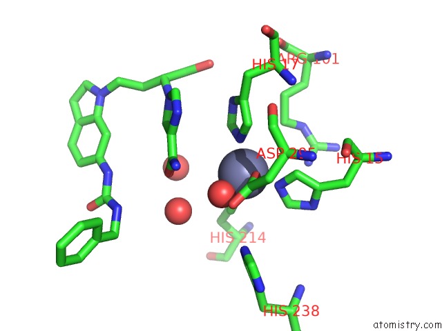
Mono view
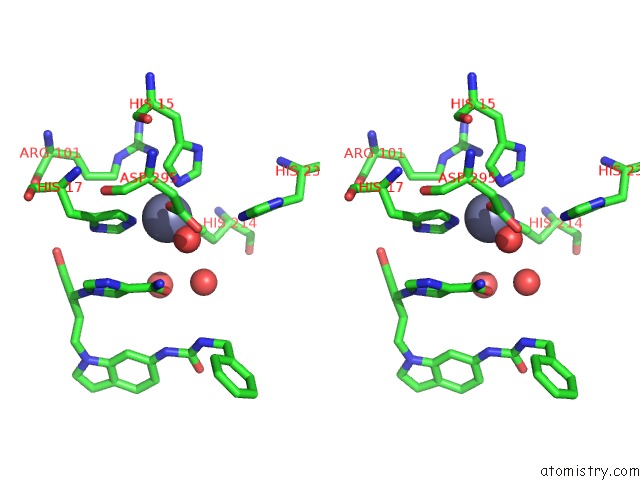
Stereo pair view
Mono view
Stereo pair view
A full contact list of Zinc with other atoms in the Zn binding
site number 1 of Crystal Structure of Adenosine Deaminase Complexed with A Potent Inhibitor within 5.0Å range:
|
Reference:
T.Terasaka,
T.Kinoshita,
M.Kuno,
N.Seki,
K.Tanaka,
I.Nakanishi.
Structure-Based Design, Synthesis, and Structure-Activity Relationship Studies of Novel Non-Nucleoside Adenosine Deaminase Inhibitors J.Med.Chem. V. 47 3730 2004.
ISSN: ISSN 0022-2623
PubMed: 15239652
DOI: 10.1021/JM0306374
Page generated: Wed Oct 16 17:28:09 2024
ISSN: ISSN 0022-2623
PubMed: 15239652
DOI: 10.1021/JM0306374
Last articles
Zn in 9J0NZn in 9J0O
Zn in 9J0P
Zn in 9FJX
Zn in 9EKB
Zn in 9C0F
Zn in 9CAH
Zn in 9CH0
Zn in 9CH3
Zn in 9CH1