Zinc »
PDB 1m2o-1ml2 »
1mea »
Zinc in PDB 1mea: Methionyl-Trna Synthetase Zinc Binding Domain. 3D Structure and Homology with Rubredoxin and Gag Retroviral Proteins
Enzymatic activity of Methionyl-Trna Synthetase Zinc Binding Domain. 3D Structure and Homology with Rubredoxin and Gag Retroviral Proteins
All present enzymatic activity of Methionyl-Trna Synthetase Zinc Binding Domain. 3D Structure and Homology with Rubredoxin and Gag Retroviral Proteins:
6.1.1.10;
6.1.1.10;
Zinc Binding Sites:
The binding sites of Zinc atom in the Methionyl-Trna Synthetase Zinc Binding Domain. 3D Structure and Homology with Rubredoxin and Gag Retroviral Proteins
(pdb code 1mea). This binding sites where shown within
5.0 Angstroms radius around Zinc atom.
In total only one binding site of Zinc was determined in the Methionyl-Trna Synthetase Zinc Binding Domain. 3D Structure and Homology with Rubredoxin and Gag Retroviral Proteins, PDB code: 1mea:
In total only one binding site of Zinc was determined in the Methionyl-Trna Synthetase Zinc Binding Domain. 3D Structure and Homology with Rubredoxin and Gag Retroviral Proteins, PDB code: 1mea:
Zinc binding site 1 out of 1 in 1mea
Go back to
Zinc binding site 1 out
of 1 in the Methionyl-Trna Synthetase Zinc Binding Domain. 3D Structure and Homology with Rubredoxin and Gag Retroviral Proteins
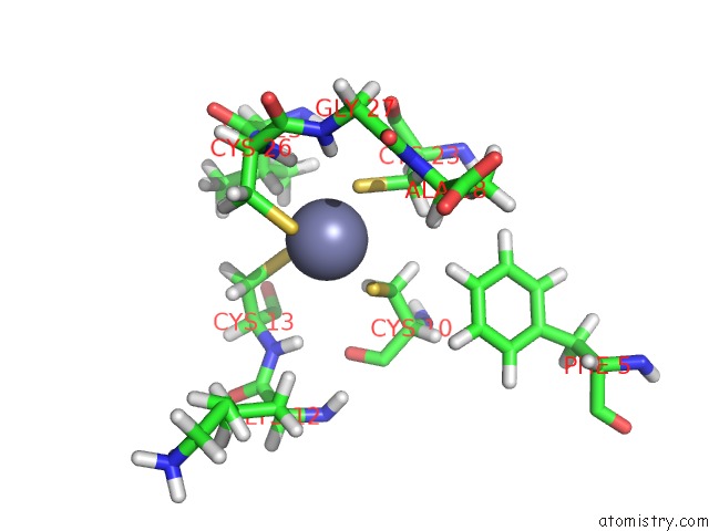
Mono view
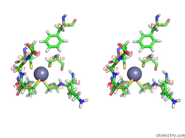
Stereo pair view
Mono view
Stereo pair view
A full contact list of Zinc with other atoms in the Zn binding
site number 1 of Methionyl-Trna Synthetase Zinc Binding Domain. 3D Structure and Homology with Rubredoxin and Gag Retroviral Proteins within 5.0Å range:
|
Reference:
D.Fourmy,
F.Dardel,
S.Blanquet.
Methionyl-Trna Synthetase Zinc Binding Domain. Three-Dimensional Structure and Homology with Rubredoxin and Gag Retroviral Proteins. J.Mol.Biol. V. 231 1078 1993.
ISSN: ISSN 0022-2836
PubMed: 8515466
DOI: 10.1006/JMBI.1993.1353
Page generated: Sun Oct 13 05:30:47 2024
ISSN: ISSN 0022-2836
PubMed: 8515466
DOI: 10.1006/JMBI.1993.1353
Last articles
Zn in 9J0NZn in 9J0O
Zn in 9J0P
Zn in 9FJX
Zn in 9EKB
Zn in 9C0F
Zn in 9CAH
Zn in 9CH0
Zn in 9CH3
Zn in 9CH1