Zinc »
PDB 1m2o-1ml2 »
1m65 »
Zinc in PDB 1m65: Ycdx Protein
Protein crystallography data
The structure of Ycdx Protein, PDB code: 1m65
was solved by
A.Teplyakov,
G.Obmolova,
P.P.Khil,
R.D.Camerini-Otero,
G.L.Gilliland,
Structure 2 Function Project (S2F),
with X-Ray Crystallography technique. A brief refinement statistics is given in the table below:
| Resolution Low / High (Å) | 10.00 / 1.57 |
| Space group | P 3 2 1 |
| Cell size a, b, c (Å), α, β, γ (°) | 77.230, 77.230, 79.990, 90.00, 90.00, 120.00 |
| R / Rfree (%) | 17.7 / 20.8 |
Other elements in 1m65:
The structure of Ycdx Protein also contains other interesting chemical elements:
| Sodium | (Na) | 3 atoms |
Zinc Binding Sites:
The binding sites of Zinc atom in the Ycdx Protein
(pdb code 1m65). This binding sites where shown within
5.0 Angstroms radius around Zinc atom.
In total only one binding site of Zinc was determined in the Ycdx Protein, PDB code: 1m65:
In total only one binding site of Zinc was determined in the Ycdx Protein, PDB code: 1m65:
Zinc binding site 1 out of 1 in 1m65
Go back to
Zinc binding site 1 out
of 1 in the Ycdx Protein
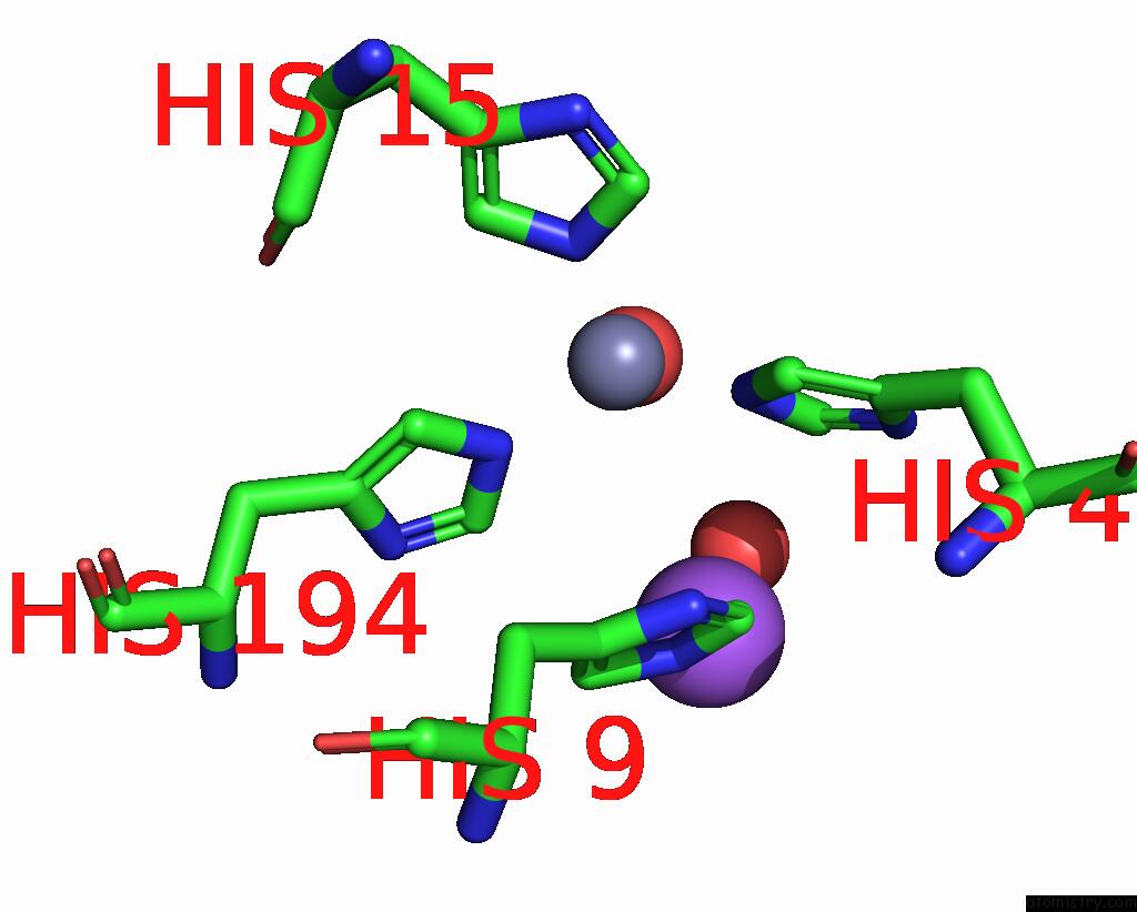
Mono view
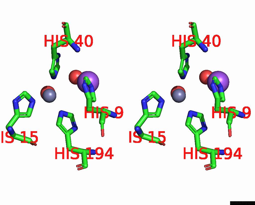
Stereo pair view
Mono view
Stereo pair view
A full contact list of Zinc with other atoms in the Zn binding
site number 1 of Ycdx Protein within 5.0Å range:
|
Reference:
A.Teplyakov,
G.Obmolova,
P.P.Khil,
A.J.Howard,
R.D.Camerini-Otero,
G.L.Gilliland.
Crystal Structure of the Escherichia Coli Ycdx Protein Reveals A Trinuclear Zinc Active Site Proteins: V. 51 315 2003STRUCT.,Funct.,Genet..
ISSN: ISSN 0887-3585
PubMed: 12661000
DOI: 10.1002/PROT.10352
Page generated: Sun Oct 13 05:26:07 2024
ISSN: ISSN 0887-3585
PubMed: 12661000
DOI: 10.1002/PROT.10352
Last articles
Zn in 9J0NZn in 9J0O
Zn in 9J0P
Zn in 9FJX
Zn in 9EKB
Zn in 9C0F
Zn in 9CAH
Zn in 9CH0
Zn in 9CH3
Zn in 9CH1