Zinc »
PDB 1m2o-1ml2 »
1m4m »
Zinc in PDB 1m4m: Mouse Survivin
Protein crystallography data
The structure of Mouse Survivin, PDB code: 1m4m
was solved by
S.W.Muchmore,
J.Chen,
C.Jakob,
D.Zakula,
E.D.Matayoshi,
W.Wu,
H.Zhang,
F.Li,
S.C.Ng,
D.C.Altieri,
with X-Ray Crystallography technique. A brief refinement statistics is given in the table below:
| Resolution Low / High (Å) | 50.00 / 2.80 |
| Space group | P 61 2 2 |
| Cell size a, b, c (Å), α, β, γ (°) | 41.430, 41.430, 291.340, 90.00, 90.00, 120.00 |
| R / Rfree (%) | n/a / n/a |
Zinc Binding Sites:
The binding sites of Zinc atom in the Mouse Survivin
(pdb code 1m4m). This binding sites where shown within
5.0 Angstroms radius around Zinc atom.
In total 2 binding sites of Zinc where determined in the Mouse Survivin, PDB code: 1m4m:
Jump to Zinc binding site number: 1; 2;
In total 2 binding sites of Zinc where determined in the Mouse Survivin, PDB code: 1m4m:
Jump to Zinc binding site number: 1; 2;
Zinc binding site 1 out of 2 in 1m4m
Go back to
Zinc binding site 1 out
of 2 in the Mouse Survivin
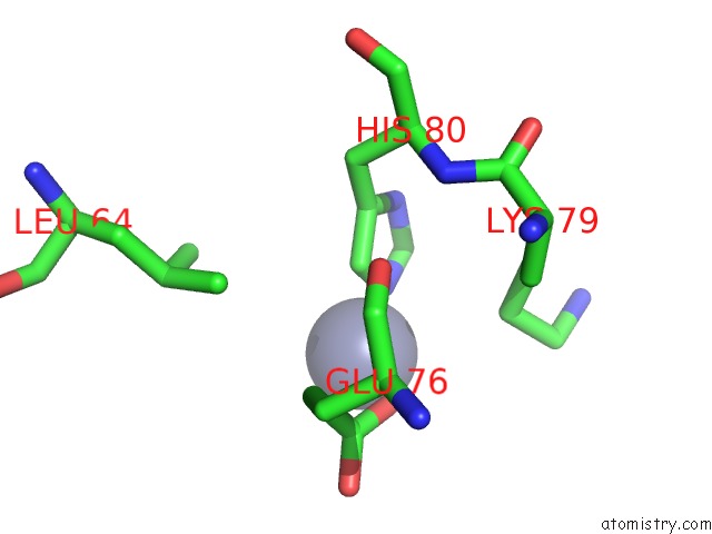
Mono view
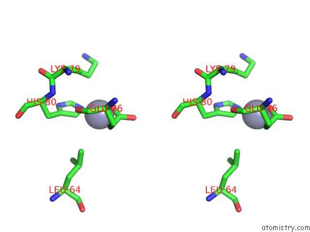
Stereo pair view
Mono view
Stereo pair view
A full contact list of Zinc with other atoms in the Zn binding
site number 1 of Mouse Survivin within 5.0Å range:
|
Zinc binding site 2 out of 2 in 1m4m
Go back to
Zinc binding site 2 out
of 2 in the Mouse Survivin
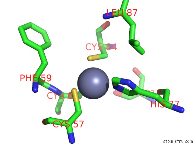
Mono view
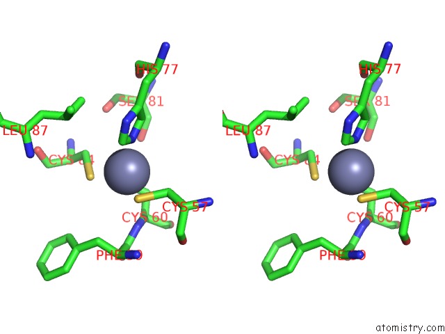
Stereo pair view
Mono view
Stereo pair view
A full contact list of Zinc with other atoms in the Zn binding
site number 2 of Mouse Survivin within 5.0Å range:
|
Reference:
S.W.Muchmore,
J.Chen,
C.Jakob,
D.Zakula,
E.D.Matayoshi,
W.Wu,
H.Zhang,
F.Li,
S.C.Ng,
D.C.Altieri.
Crystal Structure and Mutagenic Analysis of the Inhibitor-of-Apoptosis Protein Survivin Mol.Cell V. 6 173 2000.
ISSN: ISSN 1097-2765
PubMed: 10949038
DOI: 10.1016/S1097-2765(00)00018-6
Page generated: Sun Oct 13 05:19:57 2024
ISSN: ISSN 1097-2765
PubMed: 10949038
DOI: 10.1016/S1097-2765(00)00018-6
Last articles
Zn in 9J0NZn in 9J0O
Zn in 9J0P
Zn in 9FJX
Zn in 9EKB
Zn in 9C0F
Zn in 9CAH
Zn in 9CH0
Zn in 9CH3
Zn in 9CH1