Zinc »
PDB 1lgd-1m2n »
1ltx »
Zinc in PDB 1ltx: Structure of Rab Escort Protein-1 in Complex with Rab Geranylgeranyl Transferase and Isoprenoid
Protein crystallography data
The structure of Structure of Rab Escort Protein-1 in Complex with Rab Geranylgeranyl Transferase and Isoprenoid, PDB code: 1ltx
was solved by
O.Pylypenko,
A.Rak,
R.Reents,
A.Niculae,
N.H.Thoma,
H.Waldmann,
I.Schlichting,
R.S.Goody,
K.Alexandrov,
with X-Ray Crystallography technique. A brief refinement statistics is given in the table below:
| Resolution Low / High (Å) | 19.74 / 2.70 |
| Space group | P 1 21 1 |
| Cell size a, b, c (Å), α, β, γ (°) | 68.700, 197.300, 85.300, 90.00, 112.80, 90.00 |
| R / Rfree (%) | 22.4 / 27.2 |
Other elements in 1ltx:
The structure of Structure of Rab Escort Protein-1 in Complex with Rab Geranylgeranyl Transferase and Isoprenoid also contains other interesting chemical elements:
| Chlorine | (Cl) | 1 atom |
Zinc Binding Sites:
The binding sites of Zinc atom in the Structure of Rab Escort Protein-1 in Complex with Rab Geranylgeranyl Transferase and Isoprenoid
(pdb code 1ltx). This binding sites where shown within
5.0 Angstroms radius around Zinc atom.
In total only one binding site of Zinc was determined in the Structure of Rab Escort Protein-1 in Complex with Rab Geranylgeranyl Transferase and Isoprenoid, PDB code: 1ltx:
In total only one binding site of Zinc was determined in the Structure of Rab Escort Protein-1 in Complex with Rab Geranylgeranyl Transferase and Isoprenoid, PDB code: 1ltx:
Zinc binding site 1 out of 1 in 1ltx
Go back to
Zinc binding site 1 out
of 1 in the Structure of Rab Escort Protein-1 in Complex with Rab Geranylgeranyl Transferase and Isoprenoid
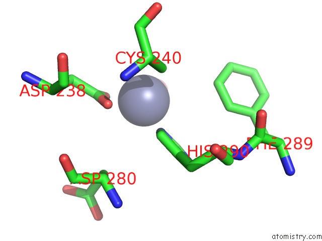
Mono view
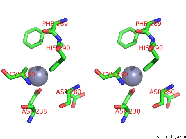
Stereo pair view
Mono view
Stereo pair view
A full contact list of Zinc with other atoms in the Zn binding
site number 1 of Structure of Rab Escort Protein-1 in Complex with Rab Geranylgeranyl Transferase and Isoprenoid within 5.0Å range:
|
Reference:
O.Pylypenko,
A.Rak,
R.Reents,
A.Niculae,
V.Sidorovitch,
M.D.Cioaca,
E.Bessolitsyna,
N.H.Thoma,
H.Waldmann,
I.Schlichting,
R.S.Goody,
K.Alexandrov.
Structure of Rab Escort Protein-1 in Complex with Rab Geranylgeranyltransferase Mol.Cell V. 11 483 2003.
ISSN: ISSN 1097-2765
PubMed: 12620235
DOI: 10.1016/S1097-2765(03)00044-3
Page generated: Tue Aug 19 21:36:35 2025
ISSN: ISSN 1097-2765
PubMed: 12620235
DOI: 10.1016/S1097-2765(03)00044-3
Last articles
Zn in 1WWGZn in 1WWE
Zn in 1WWF
Zn in 1WW1
Zn in 1WWD
Zn in 1WUR
Zn in 1WUQ
Zn in 1WUP
Zn in 1WPL
Zn in 1WUO