Zinc »
PDB 1jao-1joe »
1jmu »
Zinc in PDB 1jmu: Crystal Structure of the Reovirus MU1/SIGMA3 Complex
Protein crystallography data
The structure of Crystal Structure of the Reovirus MU1/SIGMA3 Complex, PDB code: 1jmu
was solved by
S.Liemann,
M.L.Nibert,
S.C.Harrison,
with X-Ray Crystallography technique. A brief refinement statistics is given in the table below:
| Resolution Low / High (Å) | 35.00 / 2.80 |
| Space group | I 2 2 2 |
| Cell size a, b, c (Å), α, β, γ (°) | 180.294, 184.942, 284.324, 90.00, 90.00, 90.00 |
| R / Rfree (%) | 21.6 / 24.1 |
Other elements in 1jmu:
The structure of Crystal Structure of the Reovirus MU1/SIGMA3 Complex also contains other interesting chemical elements:
| Chlorine | (Cl) | 1 atom |
Zinc Binding Sites:
The binding sites of Zinc atom in the Crystal Structure of the Reovirus MU1/SIGMA3 Complex
(pdb code 1jmu). This binding sites where shown within
5.0 Angstroms radius around Zinc atom.
In total 3 binding sites of Zinc where determined in the Crystal Structure of the Reovirus MU1/SIGMA3 Complex, PDB code: 1jmu:
Jump to Zinc binding site number: 1; 2; 3;
In total 3 binding sites of Zinc where determined in the Crystal Structure of the Reovirus MU1/SIGMA3 Complex, PDB code: 1jmu:
Jump to Zinc binding site number: 1; 2; 3;
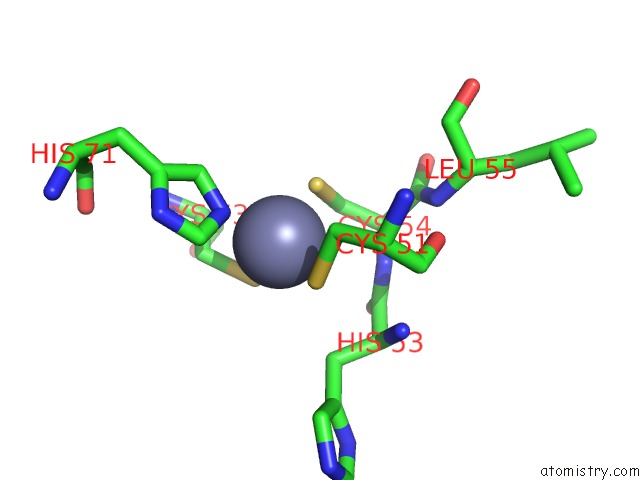
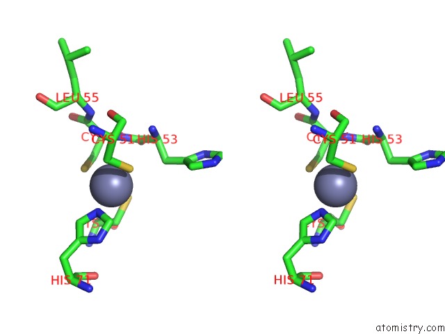
Zinc binding site 1 out of 3 in 1jmu
Go back to
Zinc binding site 1 out
of 3 in the Crystal Structure of the Reovirus MU1/SIGMA3 Complex
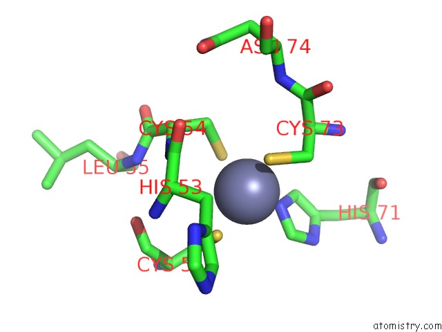
Mono view
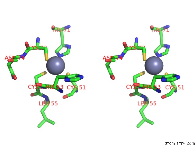
Stereo pair view
Mono view
Stereo pair view
A full contact list of Zinc with other atoms in the Zn binding
site number 1 of Crystal Structure of the Reovirus MU1/SIGMA3 Complex within 5.0Å range:
|
Zinc binding site 2 out of 3 in 1jmu
Go back to
Zinc binding site 2 out
of 3 in the Crystal Structure of the Reovirus MU1/SIGMA3 Complex
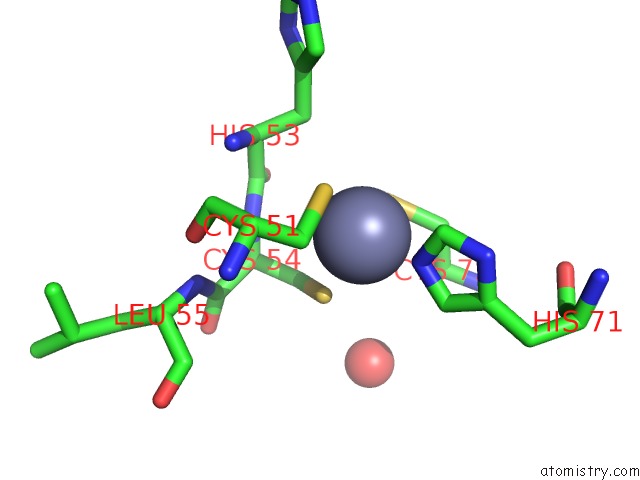
Mono view
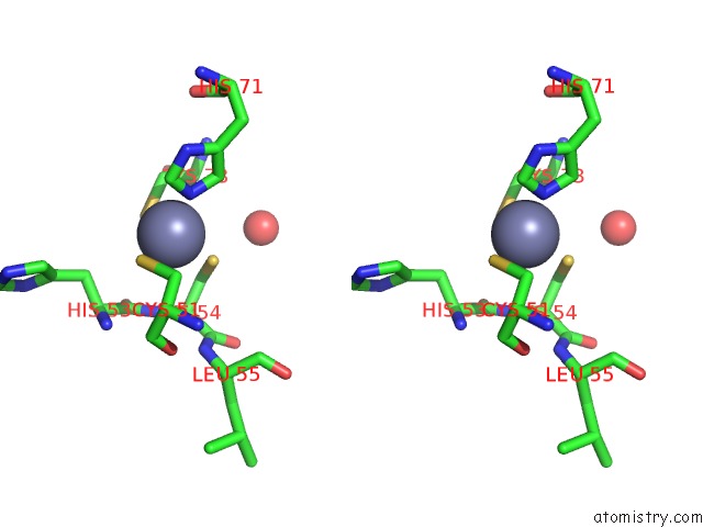
Stereo pair view
Mono view
Stereo pair view
A full contact list of Zinc with other atoms in the Zn binding
site number 2 of Crystal Structure of the Reovirus MU1/SIGMA3 Complex within 5.0Å range:
|
Zinc binding site 3 out of 3 in 1jmu
Go back to
Zinc binding site 3 out
of 3 in the Crystal Structure of the Reovirus MU1/SIGMA3 Complex
Mono view
Stereo pair view
Mono view
Stereo pair view
A full contact list of Zinc with other atoms in the Zn binding
site number 3 of Crystal Structure of the Reovirus MU1/SIGMA3 Complex within 5.0Å range:
|
Reference:
S.Liemann,
K.Chandran,
T.S.Baker,
M.L.Nibert,
S.C.Harrison.
Structure of the Reovirus Membrane-Penetration Protein, MU1, in A Complex with Is Protector Protein, SIGMA3. Cell(Cambridge,Mass.) V. 108 283 2002.
ISSN: ISSN 0092-8674
PubMed: 11832217
DOI: 10.1016/S0092-8674(02)00612-8
Page generated: Tue Aug 19 21:05:31 2025
ISSN: ISSN 0092-8674
PubMed: 11832217
DOI: 10.1016/S0092-8674(02)00612-8
Last articles
Zn in 1WWGZn in 1WWE
Zn in 1WWF
Zn in 1WW1
Zn in 1WWD
Zn in 1WUR
Zn in 1WUQ
Zn in 1WUP
Zn in 1WPL
Zn in 1WUO