Zinc »
PDB 1jao-1joe »
1jd6 »
Zinc in PDB 1jd6: Crystal Structure of DIAP1-BIR2/Hid Complex
Protein crystallography data
The structure of Crystal Structure of DIAP1-BIR2/Hid Complex, PDB code: 1jd6
was solved by
J.W.Wu,
A.E.Cocina,
J.Chai,
B.A.Hay,
Y.Shi,
with X-Ray Crystallography technique. A brief refinement statistics is given in the table below:
| Resolution Low / High (Å) | 20.00 / 2.70 |
| Space group | P 65 2 2 |
| Cell size a, b, c (Å), α, β, γ (°) | 62.700, 62.700, 130.700, 90.00, 90.00, 120.00 |
| R / Rfree (%) | 21.7 / 28.7 |
Zinc Binding Sites:
The binding sites of Zinc atom in the Crystal Structure of DIAP1-BIR2/Hid Complex
(pdb code 1jd6). This binding sites where shown within
5.0 Angstroms radius around Zinc atom.
In total only one binding site of Zinc was determined in the Crystal Structure of DIAP1-BIR2/Hid Complex, PDB code: 1jd6:
In total only one binding site of Zinc was determined in the Crystal Structure of DIAP1-BIR2/Hid Complex, PDB code: 1jd6:
Zinc binding site 1 out of 1 in 1jd6
Go back to
Zinc binding site 1 out
of 1 in the Crystal Structure of DIAP1-BIR2/Hid Complex
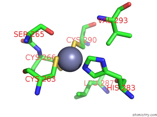
Mono view
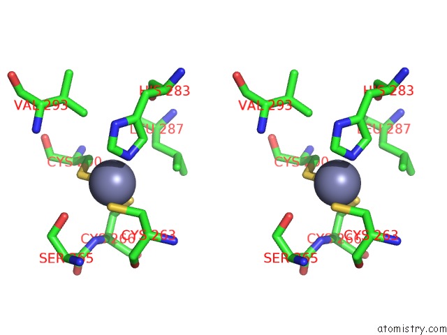
Stereo pair view
Mono view
Stereo pair view
A full contact list of Zinc with other atoms in the Zn binding
site number 1 of Crystal Structure of DIAP1-BIR2/Hid Complex within 5.0Å range:
|
Reference:
J.W.Wu,
A.E.Cocina,
J.Chai,
B.A.Hay,
Y.Shi.
Structural Analysis of A Functional DIAP1 Fragment Bound to Grim and Hid Peptides. Mol.Cell V. 8 95 2001.
ISSN: ISSN 1097-2765
PubMed: 11511363
DOI: 10.1016/S1097-2765(01)00282-9
Page generated: Sun Oct 13 03:32:05 2024
ISSN: ISSN 1097-2765
PubMed: 11511363
DOI: 10.1016/S1097-2765(01)00282-9
Last articles
Zn in 9J0NZn in 9J0O
Zn in 9J0P
Zn in 9FJX
Zn in 9EKB
Zn in 9C0F
Zn in 9CAH
Zn in 9CH0
Zn in 9CH3
Zn in 9CH1